Pentax vs. Olympus vs. Fujinon: Which Endoscopy System Is Right for Your Facility in 2026?
admin
March 23, 2026
Introduction
The three dominant manufacturers in GI endoscopy — Olympus, Pentax Medical, and Fujinon (Fujifilm) — each offer a compelling platform. Each has its flagship imaging technology, its preferred scope ecosystem, and its target facility type. And each has sales teams that will tell you theirs is the best choice.
This article takes a different approach. Rather than reproducing marketing materials, we compare the three platforms on the technical and clinical criteria that actually matter when making a capital equipment decision: imaging modality depth, scope portfolio breadth, light source technology, workflow integration, and the specific facility contexts where each system performs best.
Whether you are equipping a new GI suite, evaluating a system upgrade, or sourcing certified pre-owned endoscopy equipment, this comparison gives you the framework to make an informed decision — independent of vendor positioning.
The Three Platforms at a Glance
Before going deep into each dimension, here is a top-level overview of the flagship system from each manufacturer currently available in the U.S. and international markets as of 2026.
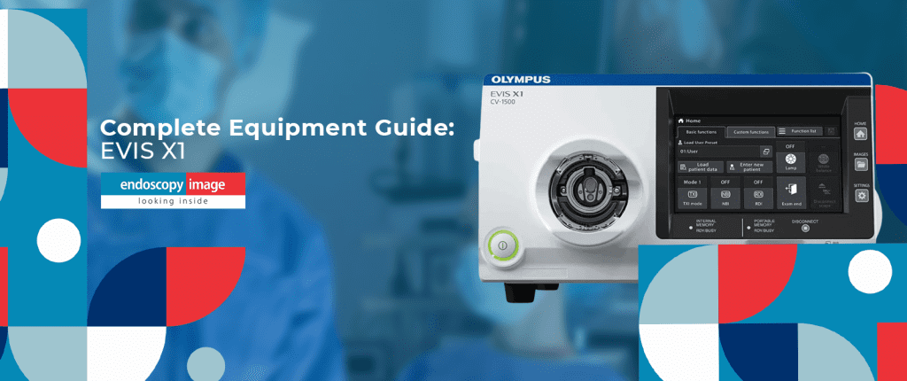
EVIS X1 — CV-1500
Olympus’s most advanced GI platform as of 2026. Flagship of the EVIS line, combining a 5-LED light engine with TXI, NBI, RDI, BAI-MAC, and the new EDOF imaging technology. FDA-cleared in 2023; EZ1500 EDOF scopes cleared May 2025.
EPK-i7010 OPTIVISTA
Pentax’s premium GI processor, featuring i-SCAN digital enhancement and OE (Optical Enhancement) technology — a hybrid digital-optical virtual chromoendoscopy platform. HD+ imaging with freeze-scan technology.
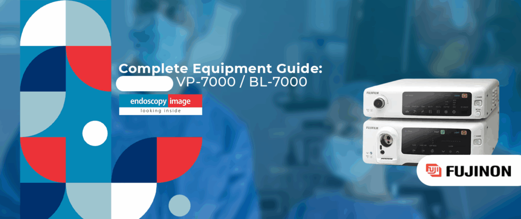
ELUXEO VP-7000 / BL-7000
Fujifilm’s LED-based ELUXEO platform, featuring 4-LED Multi-Light technology with BLI (Blue Light Imaging) and LCI (Linked Color Imaging). The first LED-based endoscopy system commercially introduced in the U.S. (2018), now in its mature generation.
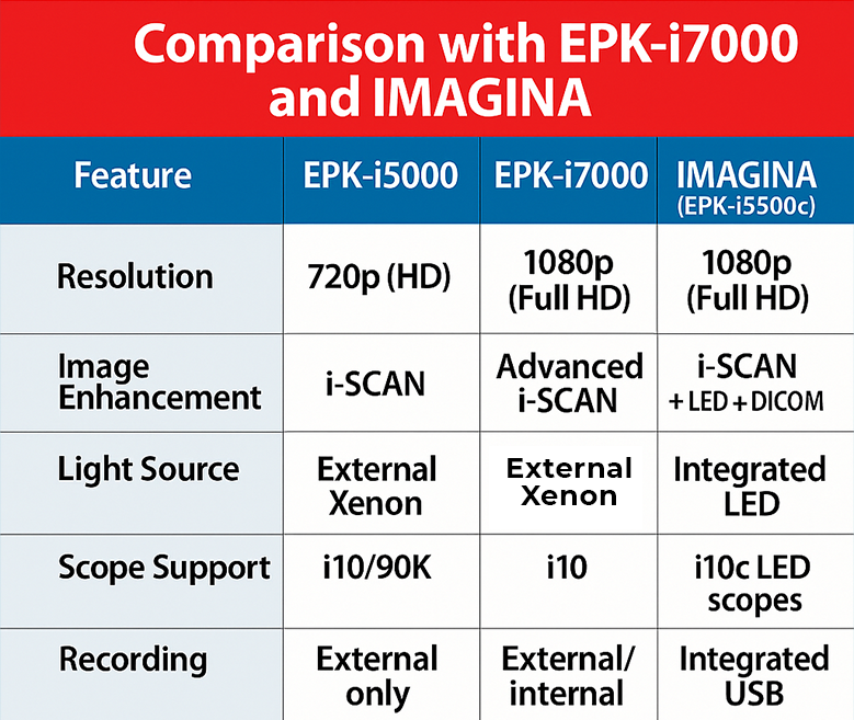
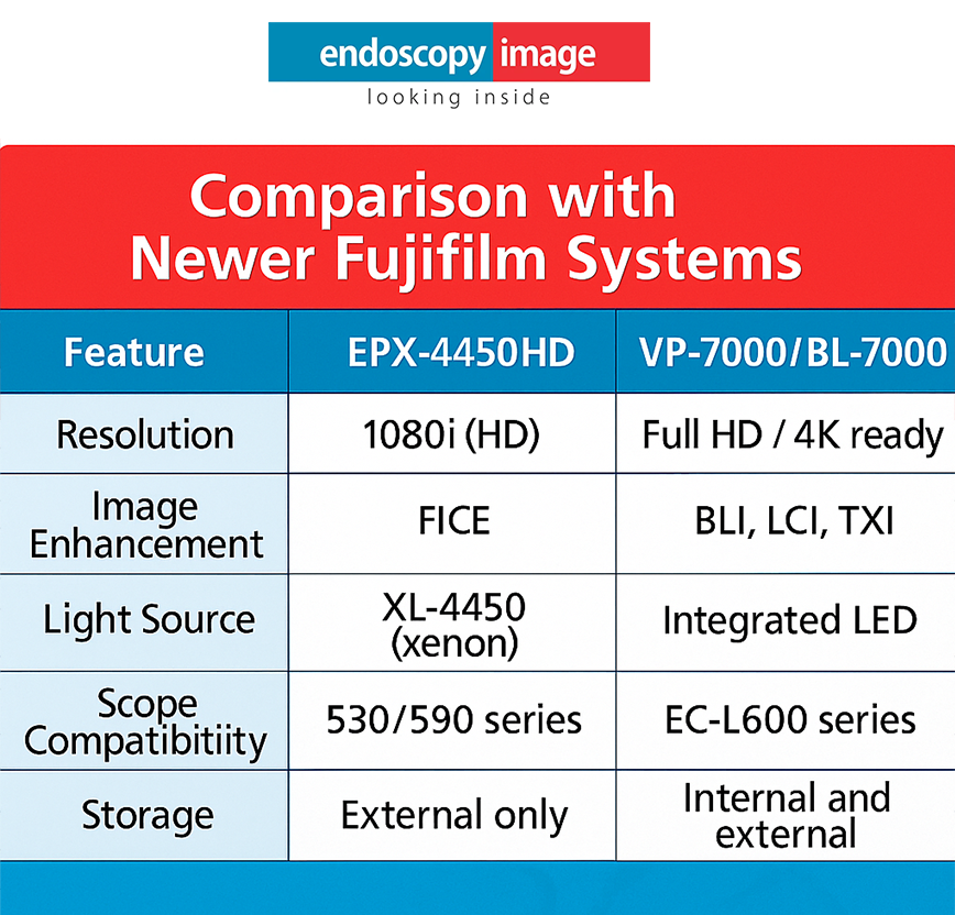
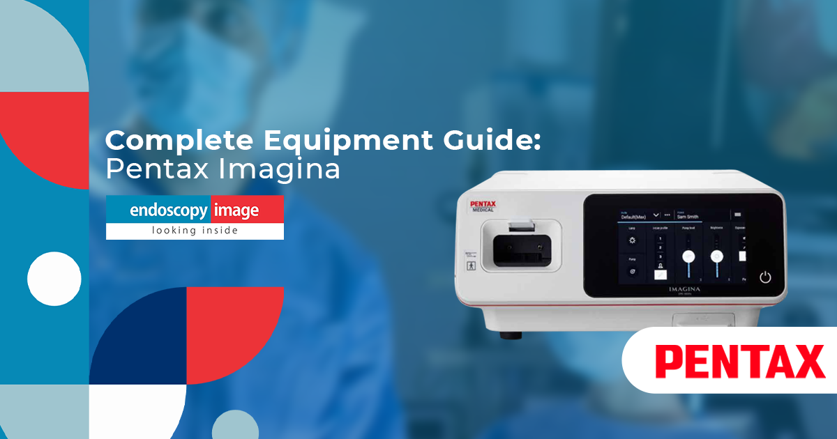
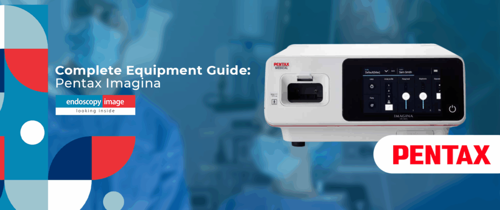
Each manufacturer also offers mid-tier or entry-level systems — Pentax’s IMAGINA, Fujinon’s EPX-4450HD (now a legacy platform), and Olympus’s EXERA III — that remain relevant for facilities with budget constraints or lower procedure volumes. These are addressed in the facility-fit section below.
Imaging Technology: How Each Brand Sees the GI Tract
The most consequential technical difference between the three platforms is their proprietary imaging enhancement technology — the modalities that go beyond standard white light endoscopy. Each brand has developed its own approach to virtual chromoendoscopy, and understanding the differences matters for clinical specialization decisions.
1. Olympus: EVIS X1 — TXI · NBI · RDI · BAI-MAC · EDOF
- NBI (Narrow Band Imaging): Olympus’s established optical-digital modality, using blue and green wavelengths absorbed by hemoglobin to enhance mucosal and vascular pattern visualization. First introduced globally in 2006; extensively published clinical evidence base.
- TXI (Texture and Color Enhancement Imaging): Digital post-processing technology designed to enhance visibility of lesions and polyps by improving color, structure, and brightness of the endoscopic image. Cleared by FDA.
- RDI (Red Dichromatic Imaging): Designed to enhance visibility of deep blood vessels and bleeding points — particularly relevant for therapeutic procedures involving GI bleeding.
- BAI-MAC (Brightness Adjustment Imaging with Maintenance of Contrast): Corrects brightness in dark areas while maintaining highlight contrast, increasing total distance view.
- EDOF (Extended Depth of Field): Olympus’s most recent innovation, using dual prisms to split light into near- and far-focused beams, producing a continuously sharp image across the full depth of field. FDA-cleared May 2025 on EZ1500 series scopes. Available in both GIF-EZ1500 gastroscope and CF-EZ1500DL/I colonoscope.
- NBI+TXI combined mode: Launched in Japan November 2025; combines NBI and TXI in a single simultaneous view. International availability pending regulatory approvals.
2. Pentax: EPK-i7010 — i-SCAN · OE Optical Enhancement · Twin Mode
- i-SCAN: Pentax’s digital image enhancement platform, combining three processing algorithms — Surface Enhancement (SE), Contrast Enhancement (CE), and Tone Enhancement (TE). Designed to provide a progressively enhanced view of mucosal texture, surface contour, and vascular structures. Software-based — no modification to the light source required.
- OE (Optical Enhancement): An optical-digital hybrid technology available on the EPK-i7010 that filters the light source to provide focused wavelength bands matching hemoglobin absorption characteristics. Combines with i-SCAN to create a three-stage examination workflow: detection, identification, and confirmation.
- Twin Mode: Displays HD+ white light and i-SCAN images side-by-side simultaneously, allowing real-time lesion characterization comparison. Designed for teaching environments and complex diagnostic sessions.
- IMAGINA i-SCAN: The IMAGINA platform includes a simplified version of i-SCAN with real-time digital enhancement, available in a more affordable system aimed at ASC and outpatient settings.
3. Fujinon: ELUXEO VP-7000 — BLI · LCI · FICE · 4-LED Multi-Light
- 4-LED Multi-Light Technology: Fujifilm’s foundational innovation for the ELUXEO platform — four independently controlled LEDs (two white, one violet, one green) that enable multiple light combinations beyond white light. First LED-based commercial GI endoscopy system introduced in the U.S. in 2018.
- BLI (Blue Light Imaging): Uses short-wavelength blue light to enhance surface structure and vascular patterns, particularly useful for lesion characterization in colonoscopy. Compatible with 700-series and selected 600-series scopes.
- LCI (Linked Color Imaging): Enhances mucosal visualization by differentiating the red color spectrum — designed to make the distinction between colorectal lesions and normal mucosa more discernible. Published clinical evidence supports its use in adenoma detection.
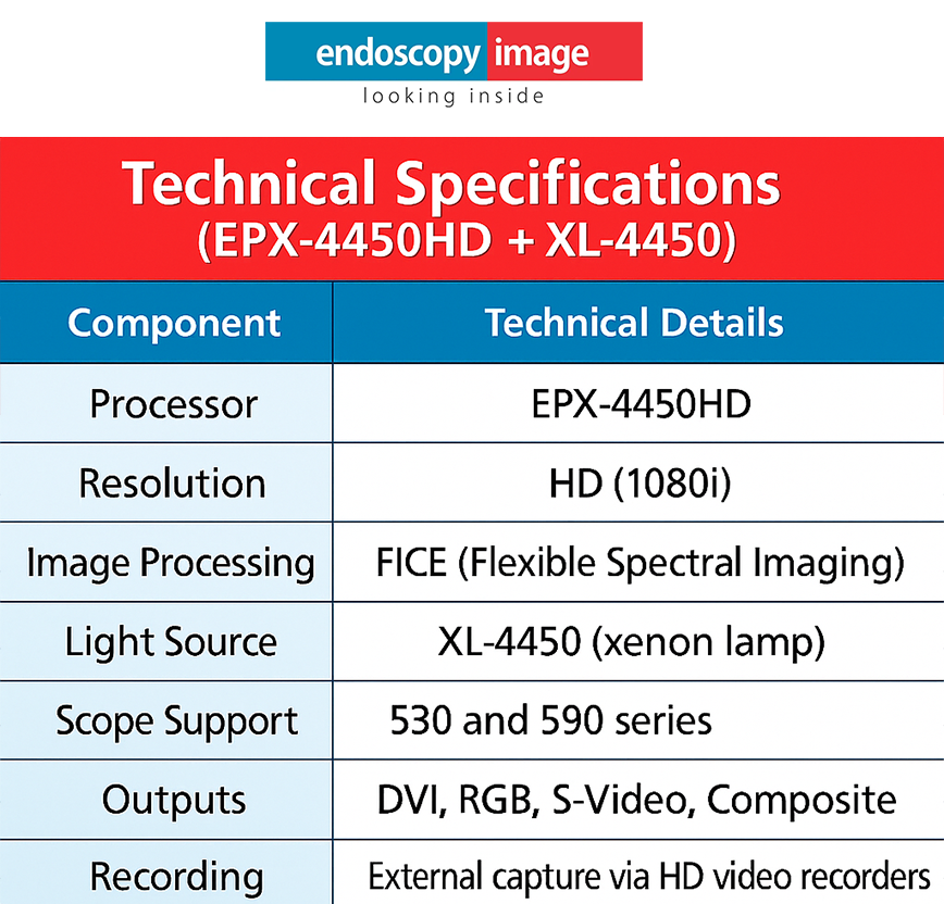
- FICE (Flexible Spectral Imaging Color Enhancement): Digital post-processing technology that allows ten configurable settings for virtual chromoendoscopy. Available on both VP-7000 and legacy EPX-4450HD platforms.
- BLI-bright mode: A modified BLI mode that increases overall brightness, improving visualization in the proximal colon and other areas with reduced luminal illumination.
Head-to-Head Comparison: Key Technical Parameters
The table below summarizes the most clinically and operationally relevant specifications across the three flagship platforms. Where a feature is platform-specific or generation-dependent, a note is included.
| Parameter | Olympus EVIS X1 | Pentax EPK-i7010 | Fujinon ELUXEO VP-7000 |
|---|---|---|---|
| Light Source | 5-LED (integrated in CV-1500) | Xenon + OE optical filter | 4-LED Multi-Light (BL-7000) |
| Image Resolution | 4K UHD (with compatible monitors) | HD+ (1080p) | Full HD |
| Proprietary Imaging | TXI, NBI, RDI, BAI-MAC, EDOF | i-SCAN, OE, Twin Mode | BLI, BLI-bright, LCI, FICE |
| AI Integration | Yes — ENDO-AID CADe (polyp detection) | Not available on EPK-i7010 currently | In development / limited availability |
| DICOM / EMR Integration | Yes | Yes (endoPRO compatible) | Yes |
| Touchscreen Interface | Yes (CV-1500 front panel) | Yes (EPK-i7010 and IMAGINA) | Partial / model-dependent |
| Scope Ergonomics | ErgoGrip — 10% lighter than prev. gen | i10c series — 20–25% lighter scopes | 700-series standard ergonomics |
| Scope Compatibility | EVIS X1 + EXERA III scopes | i10 series; limited backward compat. | 700, 600, 500 series (graded compat.) |
| Entry-level Option | EVIS EXERA III | IMAGINA (ASC-focused) | EPX-4450HD (legacy, still supported) |
| FDA Clearance (Flagship) | 2023 (EVIS X1); EDOF cleared May 2025 | FDA-cleared; year varies by model | 2017 (VP-7000 / BL-7000) |
* Resolution, AI availability, and scope compatibility may vary by configuration, region, and regulatory status. Always confirm with the manufacturer for your specific market. Information sourced from official product pages and FDA documentation as of early 2026.
NBI vs. BLI vs. i-SCAN: Understanding the Imaging Philosophy Differences
The three imaging enhancement platforms reflect fundamentally different technical philosophies — and understanding those differences helps predict which system will integrate better with your clinical workflow.
Olympus NBI: The Most Established Evidence Base
NBI was first introduced commercially in 2006 and has the largest body of peer-reviewed clinical evidence among the three optical enhancement technologies. It modifies the light source output to emit specific blue and green wavelengths that are preferentially absorbed by hemoglobin in mucosal capillaries. The result is enhanced contrast of vascular patterns and mucosal pit structures — particularly useful for Barrett’s esophagus assessment, early gastric cancer detection, and colorectal polyp characterization using validated classification systems (NICE, JNET).
The addition of TXI on the EVIS X1 platform works synergistically with NBI — and the new combined NBI+TXI mode (launched in Japan November 2025, pending international regulatory clearance) is designed to provide mucosal texture and vascular enhancement simultaneously in a single view.
Fujinon BLI and LCI: LED-Native Imaging Enhancement
Fujifilm’s approach with BLI and LCI was designed from the ground up for an LED light source — rather than adapting optical filters to a xenon system. BLI uses short-wavelength blue light to enhance surface and vascular contrast. LCI uses a different LED combination to differentiate the red color spectrum, making subtle differences between inflamed, adenomatous, and normal mucosa more visible to the endoscopist.
A meaningful body of clinical evidence has accumulated around LCI specifically in the context of colorectal adenoma detection rates. Note: while published studies have shown favorable comparisons, this is an active area of research and head-to-head comparisons with NBI are not conclusive as of 2026.
Pentax i-SCAN and OE: Digital-First Flexibility
Pentax’s i-SCAN is entirely software-based — it processes the video signal from the scope without modifying the light source. This means i-SCAN is available across Pentax’s entire compatible scope range, including older i-series instruments, without a hardware upgrade. The three-mode structure (SE, CE, TE) provides progressive examination capability — from surface detail to vascular contrast — within a single imaging pipeline.
The addition of OE (Optical Enhancement) on the EPK-i7010 brings optical filtering into the Pentax workflow for the first time, creating a hybrid digital-optical platform that narrows the gap with Olympus NBI and Fujinon BLI/LCI in terms of light-level enhancement capability. The Twin Mode — simultaneous white light and enhanced imaging on a split screen — is a genuine differentiator for training environments and complex characterization cases.
Light Source Technology: Xenon vs. LED — Why It Matters
The shift from xenon lamp-based to LED-based light sources is one of the most operationally significant transitions in endoscopy over the past decade. Both Olympus and Fujinon have moved their flagship platforms to LED-based light engines. Pentax uses a xenon source on the EPK-i7010 but integrates OE optical filtering; the IMAGINA uses distally-mounted LED lights directly in the scope tip — a different architectural approach.
| Factor | LED (Olympus EVIS X1) | LED (Fujinon VP-7000) | Xenon + OE (Pentax EPK-i7010) |
|---|---|---|---|
| Lamp replacement | Not required | Not required | Periodic replacement required |
| Warmup time | Instant | Instant | Short warmup required |
| Heat generation | Low | Low | Higher — may affect room temperature |
| Imaging mode flexibility | Multiple LED combinations enable multi-mode imaging | 4-LED independently controlled | Optical filter adds capability; fewer native modes |
| Operational cost | Lower (no bulb cost) | Lower (no bulb cost) | Ongoing lamp cost (~$300–600/replacement) |
The IMAGINA system takes a different approach: LEDs are mounted distally — at the tip of the scope itself — rather than in the processor light source. This eliminates the light-guide fiber bundle and, per Pentax’s documentation, provides more uniform illumination at the point of observation. The tradeoff is that these scopes are not backward-compatible with older Pentax processors that rely on light transmission from the proximal end.
Which System Fits Which Facility? A Practical Framework
Beyond specifications, the most useful question is: which system fits the clinical volume, case complexity, and operational constraints of your specific facility? The following framework is based on the publicly documented design intent of each platform, supplemented by the technical parameters above.
- High-volume academic and tertiary centers
- Advanced therapeutic endoscopy programs (ESD, ERCP, EUS)
- Facilities prioritizing AI-assisted polyp detection (ENDO-AID)
- Units requiring the most current imaging evidence base
- Programs conducting endoscopy research or clinical trials
- Facilities where ergonomic fatigue is a concern (ErgoGrip)
- Diagnostic and screening-focused GI units
- Ambulatory Surgery Centers (ASC) — especially IMAGINA
- Training programs that benefit from Twin Mode side-by-side imaging
- Facilities seeking lower total cost of ownership
- Units replacing older Pentax EPK systems (i-SCAN continuity)
- Budget-conscious setups that need genuine HD imaging
- Colorectal cancer screening programs (LCI adenoma detection evidence)
- Facilities replacing xenon-based Fujinon systems (scope ecosystem continuity)
- Mixed-volume hospitals and regional centers
- Units that have invested in 600/700-series Fujinon scope inventory
- Centers interested in LED-first imaging architecture
- Facilities upgrading from EPX-4450HD (direct migration path)
Scope Ecosystems and Backward Compatibility
A system purchase is not just a processor purchase — it is a commitment to a scope ecosystem. Evaluating backward compatibility and scope portfolio breadth is essential for facilities with existing instrument inventories.
- Olympus: The EVIS X1 platform unifies the previously separate EVIS EXERA and EVIS LUCERA scope lines into a single compatible portfolio. This is a significant practical advantage for mixed-inventory units. The newest EZ1500 EDOF scopes require the CV-1500 processor to unlock EDOF functionality.
- Pentax: EPK-i7010 is compatible with current i10 series scopes. The IMAGINA uses a distinct i10c scope line with distally-mounted LEDs — these are not backward-compatible with older Pentax processors. Facilities transitioning from EPK-i7000 to EPK-i7010 generally retain scope compatibility.
- Fujinon: The VP-7000 offers graded compatibility — 700-series scopes unlock the full BLI/LCI functionality; 600-series scopes offer partial compatibility (BLI/LCI available on selected models); 500-series scopes are compatible for white light and FICE imaging only. The upgrade path is clear and well-documented.
Conclusion: No Universal Winner — But a Right Choice for Your Facility
The comparison between Olympus, Pentax, and Fujinon does not produce a single winner. Each platform is technically capable of supporting high-quality GI endoscopy. What differentiates them are the specific clinical, operational, and economic parameters of your facility.
The Olympus EVIS X1 leads in imaging technology breadth, AI integration, and scope portfolio unification — making it the natural choice for high-complexity, high-volume academic and tertiary programs. The Pentax EPK-i7010 and IMAGINA offer a strong value proposition with genuine imaging enhancement capability and a lower total cost of ownership — particularly suited for ASCs, training environments, and diagnostic-focused units. The Fujinon ELUXEO VP-7000 provides a mature LED-native imaging platform with strong evidence around LCI for colorectal screening, and a clear scope upgrade path for existing Fujinon users.
For facilities evaluating equipment — new or certified pre-owned — across any of these three manufacturers, Endoscopy Image maintains a curated inventory of professionally serviced GI endoscopy systems and can support the technical evaluation process.
Frequently Asked Questions
➡️ This is not a question that can be answered definitively based on available evidence. Each platform offers clinically validated imaging enhancement modes, and image quality in practice depends on the specific scope model, the monitor used, the procedure type, and the endoscopist’s familiarity with the system. EVIS X1 currently has the largest evidence base for its individual technologies (particularly NBI), and its EDOF technology represents a genuinely new optical architecture. However, “best image quality” is not a clinically validated categorical claim for any single platform over the others.
➡️ No. Endoscope systems are proprietary — each manufacturer’s scopes use brand-specific connectors, communication protocols, and electrical interfaces. Cross-brand scope-processor combinations are not supported and would violate the IFU of both devices. Facility transitions between brands require a full scope and processor change within the new brand’s ecosystem.
➡️ None of the three manufacturers publish list prices for their endoscopy systems publicly, and pricing varies significantly by region, configuration, volume discounts, and service contract terms. As a general principle, the Pentax IMAGINA is positioned as the most cost-predictable entry for ASC and outpatient settings. The Olympus EVIS X1 and Fujinon ELUXEO VP-7000 are premium flagship systems with comparable capital investment requirements. Service contract cost and xenon lamp replacement (Pentax EPK-i7010) should be factored into total cost of ownership comparisons.
➡️ Yes — under the right conditions. A certified pre-owned Olympus, Pentax, or Fujinon system from a reputable source, with documented repair history, a passed leak test, and full functional validation, represents a clinically sound investment. The key requirements are: confirmed scope compatibility with the processor, manufacturer or certified repair facility servicing, and verification that the system supports your required imaging modes. Systems from the generation immediately preceding the current flagship — such as Olympus EXERA III, Pentax EPK-i7000, or Fujinon EPX-4450HD — remain fully functional and supported platforms for routine GI procedures.
➡️ As of early 2026, Olympus has the most commercially available AI integration through its ENDO-AID CADe (Computer-Aided Detection) platform, which provides real-time polyp detection assistance during colonoscopy. Fujifilm and Pentax have both indicated AI development roadmaps, but comprehensive commercially available AI-assisted detection systems comparable to ENDO-AID are not yet broadly deployed on the Fujinon or Pentax platforms in the U.S. market as of this writing. This is an actively evolving area.
Topics that might interest you:
Blog & Articles

Pentax vs. Olympus vs. Fujinon: Which Endoscopy System Is Right for Your Facility in 2026?
Discover the key differences between endoscopy and colonoscopy procedures. Expert guide for medical..
How Endoscope Reprocessing Affects Repair Frequency: What Every GI Manager Should Know | Endoscopy Image
Discover the key differences between endoscopy and colonoscopy procedures. Expert guide for medical..

Endoscope Leak Test: Step-by-Step Protocol for Olympus, Pentax, and Fujinon Scopes
Discover the key differences between endoscopy and colonoscopy procedures. Expert guide for medical..

Digestive Endoscopy vs. Colonoscopy: Key Differences, Indications, and What Every GI Specialist Should Know
Discover the key differences between endoscopy and colonoscopy procedures. Expert guide for medical..