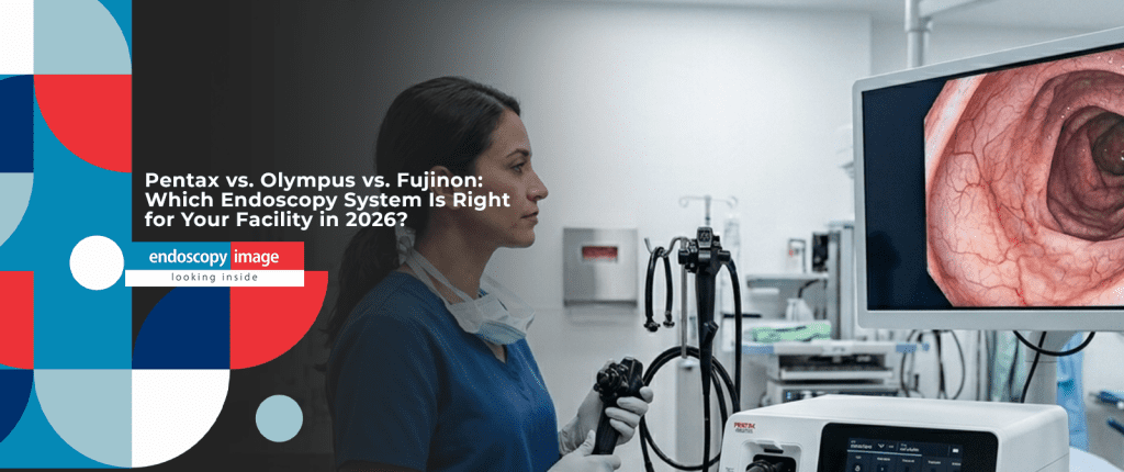
Pentax Video Processor Buying Guide: Imagina vs EPK-i5000 vs EPK-i7010
Buyer’s Guide
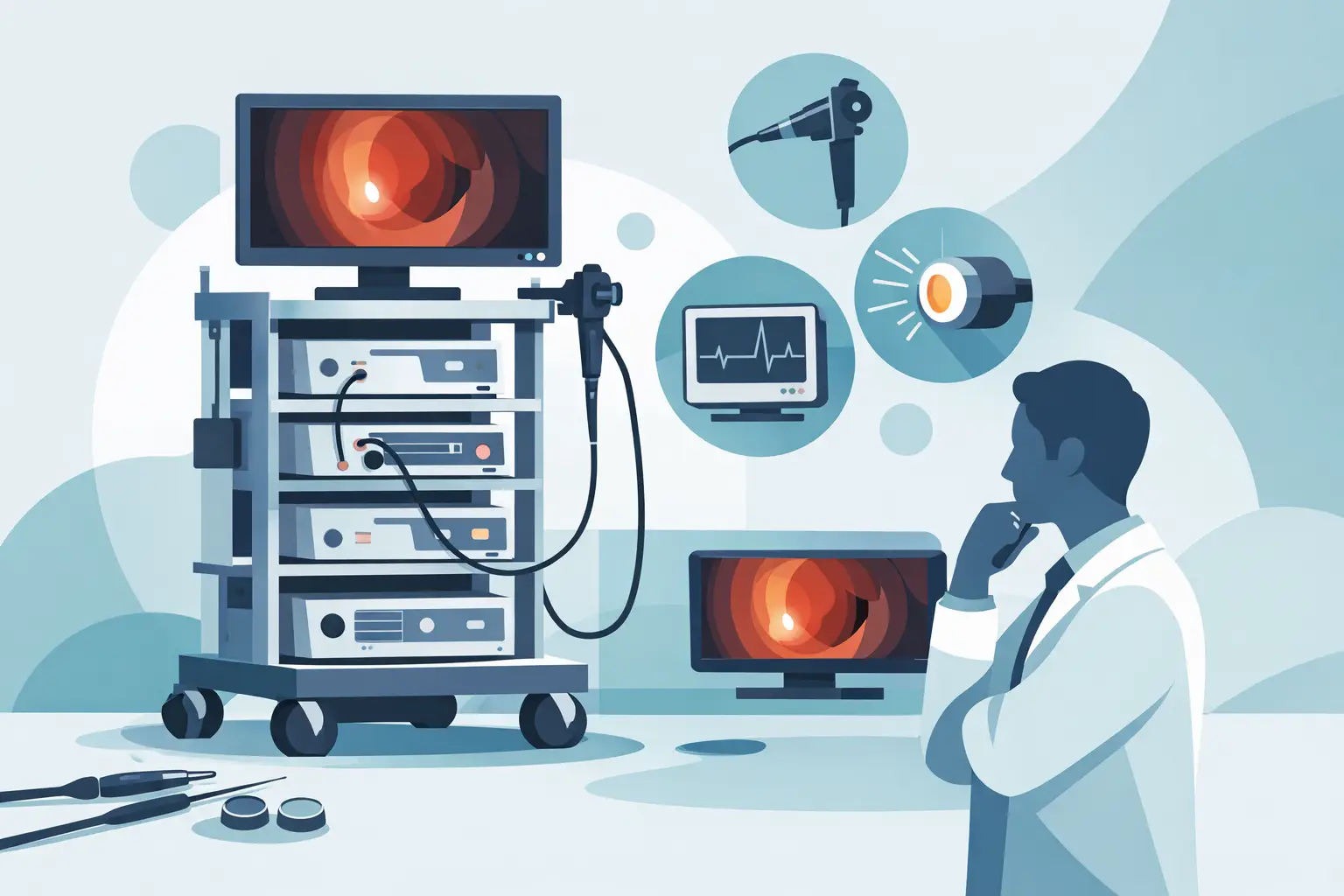
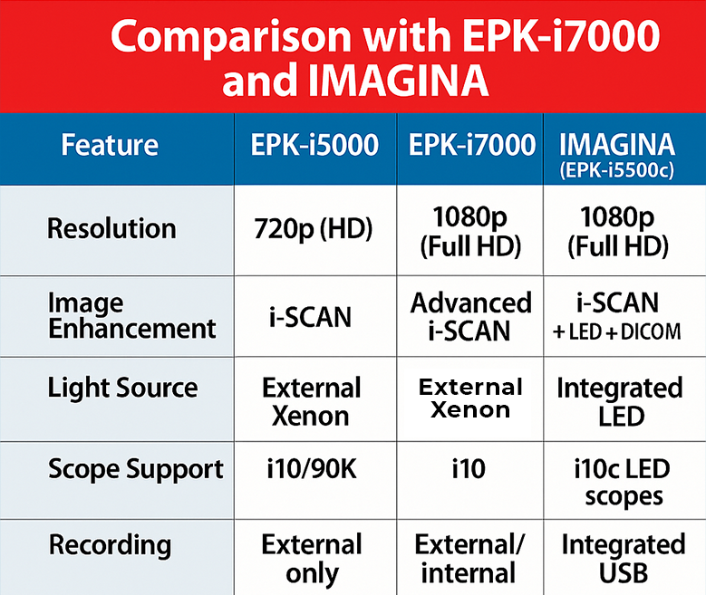
Pentax’s video processor lineup spans three clear tiers — from a compact entry system to a processor built for teaching and advanced diagnostics. Here’s how the Imagina, EPK-i5000, and OPTIVISTA EPK-i7010 actually differ, so you can match the right one to your procedure volume and clinical needs.
3 TiersEntry, mid, and advanced processors in the current lineup
i-SCANSoftware-based enhancement standard across all three
OE + TwinModeExclusive to the EPK-i7010 OPTIVISTA
20,000+ hrsLED lifespan on the Imagina, per Pentax specifications
All three Pentax processors covered here share one important trait: i-SCAN, Pentax’s proprietary image enhancement technology, is entirely software-based. It processes the video signal from the scope without changing the light source, which means it works across Pentax’s compatible scope range without requiring a hardware upgrade. That’s the foundation all three systems build on — the differences come down to light source technology, workflow features, and which additional enhancement modes are available.
Quick Comparison
Processor
Tier
Light Source
Enhancement Modes
Compatible Scopes
Pentax Imagina
Entry
Integrated LED
i-SCAN (SE/CE/TE)
i10 series
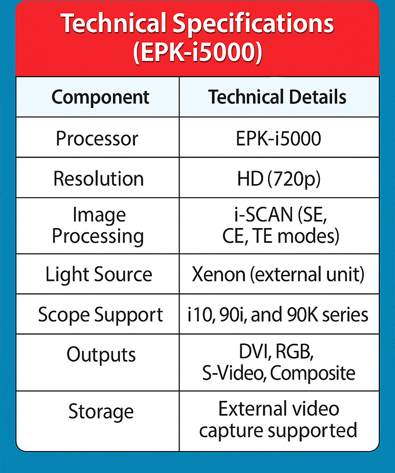
Pentax EPK-i5000
Mid
Xenon or LED
i-SCAN (SE/CE/TE)
90i, 90K series
Pentax EPK-i7010 (OPTIVISTA)
Advanced
Xenon
i-SCAN (SE/CE/TE) + OE + TwinMode
i10 HD+ series
1. Pentax Imagina — Entry-Level, Built for Routine Volume
Best For: Outpatient & Lower-Volume Clinics
The Imagina pairs an EP-HD processor with an integrated LED light source, rated by Pentax at over 20,000 hours of use with no lamp replacement needed. It supports i-SCAN’s three digital enhancement modes and connects via HDMI, LAN, and USB. It’s compact and designed for straightforward day-to-day use — gastroscopies and colonoscopies in an outpatient setting — but it’s not the right fit for complex therapeutic procedures like advanced EMR or ESD.
If your facility runs mostly routine diagnostic procedures and your priority is low maintenance overhead and a simple learning curve for staff, the Imagina is the most cost-effective entry point into Pentax’s current-generation platform.
2. Pentax EPK-i5000 — The Mid-Tier Workhorse
Best For: General GI Practices Already on Pentax
The EPK-i5000 sits between the Imagina and the EPK-i7010. It runs on either a xenon or LED light source, uses CCD sensors, and is compatible with Pentax’s 90i and 90K series scopes — a wider compatibility range if your facility has scopes from more than one generation. Like the Imagina, it relies on i-SCAN’s three modes (Surface, Contrast, and Tone Enhancement) without any optical hardware addition.
This is generally the right processor for facilities already invested in Pentax’s 90-series scope ecosystem and looking for solid, proven HD performance without moving up to the full OPTIVISTA feature set.
3. Pentax EPK-i7010 (OPTIVISTA) — Digital + Optical Enhancement
Best For: Advanced Diagnostics & Teaching Environments
The EPK-i7010, marketed by Pentax as OPTIVISTA, is the first Pentax processor to combine i-SCAN’s digital enhancement with OE (Optical Enhancement) — an optical filter that narrows the light spectrum to improve contrast on blood vessels, glandular ducts, and mucosal structure, independent of digital processing. According to Pentax Medical’s own product documentation, physicians can switch in real time between HD+ white light, i-SCAN’s digital modes, and OE — and the system’s TwinMode displays two enhancement modes side by side, which Pentax positions specifically as a teaching tool for interpreting image-enhanced endoscopy.
Why OE matters, in plain terms
i-SCAN alone works entirely through software — it doesn’t change how light hits the tissue. OE is different: it’s an optical filter that changes the light itself before the image is even captured. Combining both gives clinicians two independent ways to enhance visualization, rather than one.
This processor makes the most sense for centers doing lesion characterization work, training residents or fellows on image-enhanced endoscopy, or handling higher procedure complexity where an extra layer of visual confirmation matters.
Decision Framework: Which Tier Fits Your Facility?
Imagina
Routine, High-Volume Outpatient
Straightforward gastroscopies and colonoscopies, low maintenance overhead, budget-conscious purchase.
EPK-i5000
General GI Practice on Pentax Scopes
Already invested in 90i/90K series scopes, wants proven HD performance without the full OPTIVISTA feature set.
EPK-i7010
Advanced Diagnostics or Teaching
Lesion characterization, resident/fellow training, or procedures where optical + digital enhancement both add value.
Compatibility Snapshot
Imagina pairs with Pentax’s i10 series gastroscopes and colonoscopes.
EPK-i5000 is compatible with the 90i and 90K series, useful if your inventory spans older scopes.
EPK-i7010 works with the i10 HD+ series, and per Pentax, also supports MagniView optical zoom endoscopes.
Always confirm connector generation before buying a scope separately from the processor — even within Pentax’s own lineup, series compatibility isn’t universal.
Why a Pre-Owned Pentax System Is a Practical Way In
All three processors covered here — Imagina, EPK-i5000, and EPK-i7010 — are realistic options on the pre-owned market, which matters because it lets a clinic match its budget to the tier it actually needs, rather than defaulting to whichever new processor a distributor pushes hardest. At endoscopyimage.com, every pre-owned Pentax system is functionally tested before listing. If you want more detail on a specific model, our complete guides to the Pentax Imagina and the Pentax EPK-i5000 go deeper into each system.
i-SCAN is a digital, software-based enhancement that processes the video signal without changing the light source, so it works across Pentax’s compatible scope range without a hardware upgrade. OE (Optical Enhancement), available only on the EPK-i7010, is an optical filter that changes the light itself before capture. The EPK-i7010 is the only processor in this lineup that offers both.
Can I use my existing Pentax scopes with any of these processors?
It depends on the scope series. The Imagina pairs with i10 series scopes, the EPK-i5000 with 90i and 90K series, and the EPK-i7010 with the i10 HD+ series. Confirm your specific scope model’s compatibility before purchasing a processor separately from your scope inventory.
Is the EPK-i7010 worth it if I don’t do advanced therapeutic procedures?
Not necessarily. TwinMode and OE are most valuable for lesion characterization, teaching, and higher-complexity diagnostic work. For routine screening and general GI practice, the Imagina or EPK-i5000 typically deliver the imaging quality needed without the added cost of the OPTIVISTA feature set.
Is pre-owned Pentax equipment a reliable option?
Pre-owned Pentax equipment can be a reliable option when it has been functionally tested and comes with documentation from the seller. Endoscopy Image labels all Pentax equipment as pre-owned and tests functionality before listing.
Not Sure Which Pentax Processor Fits Your Clinic?
Tell us your procedure mix and current scope inventory — we’ll help you match the right tier, whether that’s the Imagina, the EPK-i5000, or the EPK-i7010.
5 Signs Your Fujinon Endoscopy System Needs an Upgrade
5 Signs Your Fujinon Endoscopy System Needs an Upgrade
Buyer’s Guide
Every Fujinon video processor eventually reaches a point where repairs, image quality, or missing diagnostic modes start limiting what your clinic can offer. Here’s how to recognize that point before it becomes a patient-safety or revenue problem — and what your realistic upgrade paths look like.
LCI · BLIImaging modes on the ELUXEO VP-7000 platform
LEDMulti-light source replacing xenon on ELUXEO systems
500–700Fujinon scope series compatible with ELUXEO VP-7000
ISO 13485Standard used by accredited endoscope repair facilities
Fujinon endoscopy systems — video processors like the EPX-4450HD or the ELUXEO VP-7000, paired with their compatible gastroscopes and colonoscopes — are built for years of clinical service. But “built to last” doesn’t mean “never needs replacing.” The question most clinic owners and GI department heads actually face isn’t if they’ll eventually upgrade, but when the math tips from “keep repairing” to “time to replace.”
This guide walks through the practical signals worth watching, grounded in how the industry itself talks about endoscope lifecycle, reprocessing risk, and platform obsolescence — not guesswork.
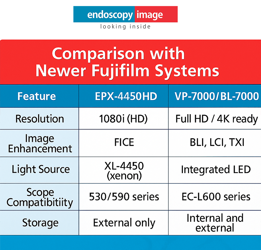
1. Image Quality No Longer Meets What Referring Physicians and Patients Expect
Clinical Signal
Endoscopic imaging technology has moved quickly. Fujifilm’s current-generation ELUXEO platform — the VP-7000/BL-7000 system, already available pre-owned through Endoscopy Image — brought Linked Color Imaging (LCI) and Blue Light Imaging (BLI) to the Fujinon lineup, both designed to improve visualization of mucosal and vascular detail during diagnostic procedures. Fujifilm’s roadmap has since moved further, with a newer ELUXEO 8000 platform announced in late 2025 — a sign of where the technology is headed, even though that specific generation is still too new to appear on the pre-owned market.
If your current Fujinon system is still running standard-definition or early HD imaging without LCI/BLI-class enhancement, that’s not a cosmetic gap — it directly affects polyp and lesion detection during procedures. When physicians in your network start comparing your equipment unfavorably to what they see at other facilities, that’s a concrete signal, not just a preference issue.
2. Repairs Are Becoming Frequent, Not Occasional
Operational Signal
What the industry says about endoscope lifespan
Flexible endoscope longevity varies enormously by handling and reprocessing discipline. As one endoscopy technology assessment expert put it in an industry interview on common endoscope handling mistakes, well-maintained scopes can remain in service well over ten years, while equipment in high-turnover or heavily used settings may need major repairs every few years. This applies broadly across endoscope brands, Fujinon included — it isn’t a brand-specific figure.
The practical takeaway: if your Fujinon processor or scopes are going in for repair multiple times within the same year — and each repair addresses a different component — that pattern usually means the system is nearing the end of its practical service life, not just having a run of bad luck. Track your service tickets. A rising frequency, even without a big single failure, is the earliest reliable warning sign.
3. Reprocessing and Compliance Risk Is Creeping Up
Compliance Signal
Reprocessing risk isn’t just about the endoscope’s age — it’s about how well the system’s design and documentation support consistent, validated cleaning. ECRI’s 2026 Health Technology Hazards report flags variability in manufacturer reprocessing instructions as an ongoing patient-safety concern, and specifically recommends that healthcare organizations assess reprocessing instructions for use (IFUs) as part of any purchasing decision — not just for new equipment, but as an ongoing risk-management practice for equipment already in service.
When to escalate this internally
If your infection control or sterile processing team has flagged repeated difficulty achieving a validated clean on a specific scope or processor — regardless of brand — that’s a patient-safety conversation, not just a maintenance one. Involve biomed and infection prevention before deciding whether to repair or replace.
4. New Fujinon Scopes No Longer Match Your Older Processor
Compatibility Signal
One detail worth understanding before you buy a replacement scope for an older Fujinon processor: Fujifilm has been moving toward broader cross-generation compatibility with its ELUXEO platforms. The VP-7000/BL-7000 system, for example, works across Fujinon’s 500-, 600-, and 700-series scopes — a real advantage if your clinic has scopes from more than one generation. Fujifilm has extended this approach further with the newer ELUXEO 8000, though that platform isn’t yet part of the pre-owned market.
What’s actually available today
For clinics looking to close a compatibility or imaging gap now, the ELUXEO VP-7000/BL-7000 is the practical, currently-available pre-owned option — not the newly released ELUXEO 8000, which hasn’t reached the secondary market yet.
If you’re finding that newer Fujinon-compatible scopes on the market won’t pair cleanly with your existing processor’s software or connector generation, that’s a sign your processor — not necessarily your scopes — is the bottleneck. In that scenario, upgrading the processor alone (rather than the full stack) is often the more cost-effective path.
5. You’re Missing Diagnostic Modes Your Peers Already Have
Competitive Signal
Systems still on standard xenon light sources
Older platforms rely on xenon lamps, which have a finite bulb life and don’t support LED-based multi-light imaging modes like LCI and BLI.
Current LED multi-light platforms
Fujifilm’s ELUXEO line uses independently regulated LED light sources, which the manufacturer states improves illumination consistency and enables imaging modes like LCI and BLI — already available on the VP-7000/BL-7000 system.
If competing clinics or hospitals in your referral network are already promoting LCI or BLI-enhanced procedures and yours isn’t, that’s both a clinical and a marketing gap worth quantifying internally before it affects referral volume.
Decision Framework: Keep, Repair, or Upgrade?
Keep
Keep As-Is
Repairs are rare and isolated, image quality still meets clinical needs, and reprocessing passes validation consistently.
Repair
Repair & Monitor
One component is failing, but the rest of the system is sound. Fix it, but start budgeting for replacement within the next cycle.
Upgrade
Time to Upgrade
Multiple repairs per year, compatibility gaps with new scopes, or missing imaging modes that affect diagnostic capability.
Why a Pre-Owned Fujinon System Is Often the Responsible Upgrade Path
Replacing an entire endoscopy stack with brand-new equipment isn’t always financially realistic, especially for independent clinics and ambulatory centers. A carefully inspected, pre-owned Fujinon system — sourced from a supplier that tests functionality and provides documentation before sale — lets you move to a more capable, better-supported platform without the full cost of new equipment.
Ask for functional testing records before purchase, not just a visual inspection.
Confirm scope-to-processor compatibility explicitly, especially across series generations.
Verify what imaging modes are active — some LCI/BLI-capable processors are sold without the full feature set enabled.
Request post-sale technical support terms in writing before committing.
At endoscopyimage.com, every pre-owned Fujinon system goes through functional testing before it’s listed. If you want to see what a current-generation, currently-available Fujinon platform looks like in detail, our complete guide to the ELUXEO VP-7000/BL-7000 system is a good next stop. Explore current inventory across our full endoscopy equipment catalog, or go directly to video gastroscopes and colonoscopy equipment if you already know which component needs replacing. If your upgrade also touches your surgical stack, our video surgery equipment catalog covers that side of the department too.
Frequently Asked Questions
How long does a Fujinon endoscopy system typically last?
There’s no fixed number that applies to every facility — flexible endoscope lifespan depends heavily on handling, reprocessing discipline, and usage volume, and this holds true across brands, not just Fujinon. Well-maintained systems can remain clinically usable for many years, while heavily used or poorly maintained equipment may need major repairs much sooner.
Is pre-owned Fujinon equipment reliable for clinical use?
Pre-owned Fujinon equipment can be a reliable option when it has been functionally tested and comes with documentation from the seller. As with any pre-owned medical device, the key is verifying test records and post-sale support terms before purchase — Endoscopy Image labels all Fujinon equipment as pre-owned and provides functional testing prior to sale.
What’s the difference between older Fujinon processors and the current ELUXEO platform?
The ELUXEO VP-7000/BL-7000, currently available pre-owned through Endoscopy Image, uses LED multi-light technology instead of xenon lamps and supports imaging modes like LCI and BLI that older Fujinon processors lack. Fujifilm has continued developing the ELUXEO line since then, but those newer releases haven’t reached the pre-owned market yet.
Should I replace my processor and scopes at the same time?
Not necessarily. If your processor is the limiting factor — for example, it can’t support newer scope generations or lacks LCI/BLI imaging — upgrading the processor alone is often more cost-effective than replacing the entire stack, provided compatibility is confirmed first.
Not Sure Where Your Fujinon System Stands?
Talk to our team about your current setup — we’ll help you figure out whether a repair, a partial upgrade, or a full pre-owned system replacement makes the most sense for your clinic.
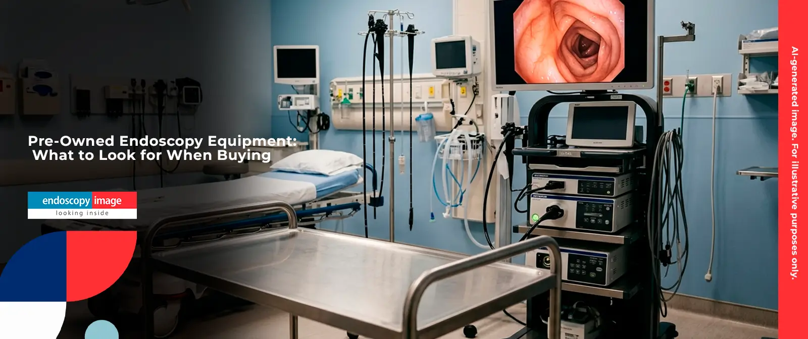
Pre-Owned Endoscopy Equipment: What to Look for When Buying
Hospitals, clinics, and surgical centers across the Americas are increasingly turning to pre-owned endoscopy systems to expand their diagnostic capacity without the full cost of new equipment. This guide walks you through what to evaluate before purchasing — and which systems are worth considering.
30+Years in the Medical Equipment Sector
7,000+Clinics & Hospitals Served Worldwide
660+Installations Completed in 2025
15+Years of U.S.-Based Operation
The market for pre-owned endoscopy equipment has grown steadily as healthcare facilities look for ways to balance quality care with budget realities. Whether you are outfitting a new clinic, replacing aging equipment, or scaling a surgical center, understanding what to look for in a pre-owned system can mean the difference between a solid investment and a costly mistake.
At Endoscopy Image, we have been operating in this space for over 30 years, serving hospitals and clinics across North America, Latin America, and beyond. This guide is designed to give procurement professionals, physicians, and clinic administrators a clear framework for evaluating pre-owned endoscopy systems — before committing to a purchase.
ℹ️ About this guide
All equipment referenced in this article is sourced from the Endoscopy Image catalog. We do not make performance claims beyond what is documented for each system. For clinical validation or technical specifications, always consult your qualified biomedical engineer.
Section 01
Why Facilities Choose Pre-Owned Endoscopy Systems
The decision to purchase pre-owned endoscopy equipment is rarely made out of compromise. For most healthcare administrators, it is a deliberate strategic choice driven by a combination of factors: capital budget constraints, faster deployment timelines, and access to proven platforms that still deliver clinically reliable image quality.
New endoscopy systems — particularly flagship platforms from leading manufacturers — carry significant acquisition costs. Pre-owned systems allow facilities to acquire equivalent diagnostic capability at a fraction of that investment, freeing budget for staffing, facility upgrades, or expanded patient capacity.
There is also a practical advantage: pre-owned platforms that have been in clinical use for several years have a well-established track record. Service documentation, known failure modes, and widely available replacement parts all contribute to lower total cost of ownership over time.
This dynamic is especially visible in markets across Latin America, Brazil, and India, where healthcare budgets are under consistent pressure and access to pre-owned medical equipment through reliable international suppliers fills a critical gap in equipment availability.
✔ Key Takeaway
Pre-owned endoscopy equipment is not a fallback — it is a deliberate procurement strategy used by hospitals and surgical centers across the Americas to optimize capital allocation while maintaining diagnostic quality.
Section 02
Understanding the Difference: Pre-Owned vs. Refurbished
Before evaluating any specific system, it helps to understand the terminology — because it affects what you should expect from the equipment and from the supplier.
Pre-owned refers to equipment that has been previously used in a clinical setting and is being resold through a secondary market channel. The term does not carry a specific implication about the extent of any reconditioning — it describes the equipment’s history of use.
Refurbished typically implies a more formal process: inspection, component-level testing, repair or replacement of worn parts, and some form of recertification before resale. In the endoscopy market, this term is most consistently associated with Olympus equipment, where the volume and depth of service infrastructure supports a meaningful reconditioning process.
⚠️ Important Distinction
Not all pre-owned equipment has undergone the same level of inspection or preparation. Always ask your supplier what, specifically, was done to the equipment before it was made available for sale — and verify through documentation.
At Endoscopy Image, equipment sold through our catalog consists of pre-owned products acquired through lawful secondary market channels. Olympus systems we carry are described as refurbished; systems from Pentax and Fujinon are offered as pre-owned. In all cases, we encourage buyers to conduct their own due diligence and involve qualified biomedical engineering staff before deployment.
Section 03
What to Evaluate Before Buying Pre-Owned Endoscopy Equipment
Purchasing a pre-owned endoscopy system requires a structured evaluation process. The following steps reflect what experienced procurement teams typically use when sourcing equipment through secondary market channels.
1
Confirm Platform Compatibility
Endoscopy processors, light sources, and scopes must be compatible within the same platform. An Olympus CV-190 processor, for example, is designed to work with the EVIS EXERA III scope series. Mixing components from different generations or platforms can result in degraded image quality or functional incompatibility. Always verify that the complete system — processor, light source, and scope — is matched before purchase.
2
Request Service and Usage History
A reputable supplier should be able to provide documentation on the equipment’s service history, including any repairs performed and the number of procedures logged where available. Equipment with a documented service record provides a clearer picture of its remaining useful life and any areas that may require attention after acquisition.
3
Assess Scope Condition Carefully
Among all components of an endoscopy system, the flexible scope (gastroscope or colonoscope) typically carries the highest risk of wear-related issues. Bending sections, insertion tubes, and optical components all degrade over time with use and reprocessing. Physical inspection by a qualified technician before deployment is strongly recommended for any pre-owned scope.
4
Verify Parts and Service Availability
Before committing to a platform, confirm that replacement parts and qualified service technicians are available in your region. Established platforms from major manufacturers typically have broader service infrastructure than less common models — a meaningful practical advantage when equipment requires maintenance.
5
Involve Your Biomedical Engineering Team
Final acceptance of any pre-owned endoscopy system should involve your facility’s biomedical engineering staff or an external qualified technician. Electrical safety testing, functional verification, and image quality assessment are standard steps before a system enters clinical use — regardless of the source of the equipment.
Section 04
Pre-Owned Endoscopy Systems Available at Endoscopy Image
The following section covers the main endoscopy platforms available through Endoscopy Image’s equipment catalog. All systems are sourced through lawful secondary market channels and shipped worldwide from our base in Key Biscayne, Miami, Florida.
Olympus — Refurbished Endoscopy Systems
Olympus is the most widely recognized name in digestive endoscopy, and their systems represent the largest segment of the refurbished endoscopy equipment market. The breadth of the Olympus installed base worldwide means that refurbished Olympus systems are relatively available and that service infrastructure is well-established in most markets.
Olympus
Refurbished
EVIS EXERA III — CV-190 / CLV-190
The CV-190 is the central video processor of the EVIS EXERA III platform. It delivers high-definition imaging with NBI (Narrow Band Imaging) technology, which enhances visualization of vascular and mucosal structures. Compatible with the 190 series scope line, including the GIF-H190 gastroscope and CF-H190 colonoscope.
Olympus
Refurbished
EVIS EXERA II — CV-170 System
The CV-170 belongs to the EVIS EXERA II platform and delivers reliable high-definition imaging for upper and lower digestive endoscopy. When paired with the GIF-H170 gastroscope and CF-H170 colonoscope, it provides a complete solution for facilities seeking a cost-effective refurbished Olympus system with proven clinical reliability.
Olympus
Refurbished
EVIS X1 — Advanced Platform
The EVIS X1 is Olympus’s most advanced endoscopy platform, featuring TXI (Texture and Color Enhancement Imaging), RDI (Red Dichromatic Imaging), NBI, and 4K UHD resolution with AI-assisted lesion detection support. Available as refurbished equipment for facilities requiring the most current Olympus imaging capability.
ℹ️ Scope Compatibility Note
Each Olympus platform (EVIS EXERA II, EVIS EXERA III, EVIS X1) uses a specific scope series. When purchasing a refurbished Olympus system, always confirm that the processor and scopes are from the same compatible generation. Mixed-platform configurations are not recommended.
Pentax — Pre-Owned Endoscopy Systems
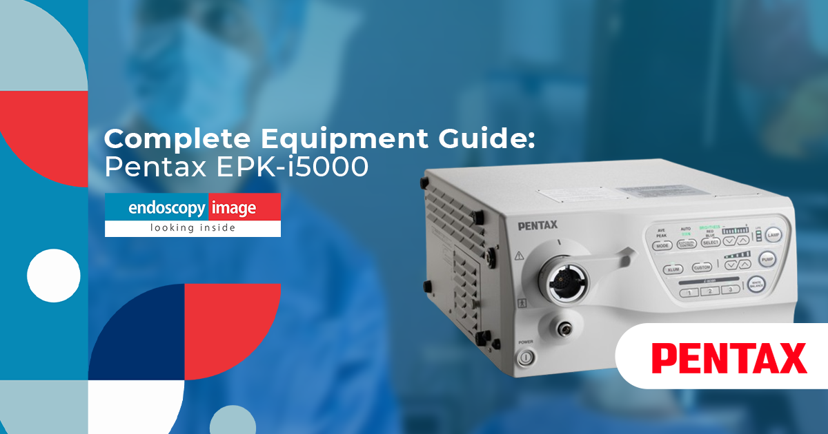
Pentax Medical produces a well-regarded line of video endoscopy processors and scopes used widely in digestive endoscopy. Their systems are available as pre-owned equipment and represent a solid alternative for facilities that need reliable HD imaging with broad scope compatibility.
Pentax
Pre-Owned
EPK-1000 System
The EPK-1000 processor, paired with the EG-2970K gastroscope and EC-3872LK colonoscope, forms a complete and robust system for upper and lower digestive endoscopy. Well-regarded for its reliability and availability in the secondary market, it is a practical option for facilities prioritizing cost-effectiveness.
Pentax
Pre-Owned
EPKi / EPKi-5010 / EPKi-7010
The EPKi line offers Full HD imaging with digital processing. The EPKi-7010, combined with the EG-29i10 gastroscope and EC-38i10L colonoscope, delivers advanced illumination and enhanced contrast for detailed mucosal visualization. The EPKi-5010 represents a consolidated cost-effective option within the same scope series.
Fujinon — Pre-Owned Endoscopy Systems
Fujinon (Fujifilm) endoscopy systems are recognized for their imaging technologies and are available as pre-owned equipment at Endoscopy Image. Their ELUXEO platform is among the more frequently requested systems in the pre-owned market.
Fujinon
Pre-Owned
ELUXEO 7000 — VP-7000 / BL-7000
The Fujinon ELUXEO 7000 system, composed of the VP-7000 processor and BL-7000 light source, offers HD imaging with LCI (Linked Color Imaging) and BLI (Blue Light Imaging) technologies. When paired with the EG-760R gastroscope and EC-760VLR colonoscope, it provides a complete solution for upper and lower digestive endoscopy.
Section 05
Platform Comparison Overview
The table below summarizes the main pre-owned and refurbished endoscopy platforms available through Endoscopy Image, to assist procurement teams in comparing options at a glance.
Brand
Platform / Processor
Resolution
Condition
Scope Series
Olympus
EVIS X1
4K UHD
Refurbished
EVIS X1 Line
Olympus
EVIS EXERA III — CV-190
Full HD
Refurbished
GIF-H190 / CF-H190
Olympus
EVIS EXERA II — CV-170
HD
Refurbished
GIF-H170 / CF-H170
Pentax
EPKi-7010
Full HD
Pre-Owned
EG-29i10 / EC-38i10L
Pentax
EPKi-5010
Full HD
Pre-Owned
EG-29i10 / EC-38i10L
Pentax
EPK-1000
HD
Pre-Owned
EG-2970K / EC-3872LK
Fujinon
ELUXEO 7000 — VP-7000
HD
Pre-Owned
EG-760R / EC-760VLR
Section 06
Tips for a Successful Pre-Owned Equipment Purchase
Beyond the technical evaluation, there are practical considerations that consistently separate successful pre-owned equipment purchases from problematic ones. The following tips are drawn from our experience working with healthcare facilities across the Americas.
🔍
Buy complete systems, not isolated components
Purchasing a processor without matching scopes — or vice versa — significantly increases compatibility risk. When possible, source the complete system (processor, light source, scopes) from a single supplier who can verify compatibility.
📋
Ask for documentation, not just assurances
Any reputable supplier should be able to provide written documentation on the equipment’s origin and condition. Verbal assurances without supporting documentation should be treated with caution.
🌎
Confirm international shipping and import compliance
Pre-owned medical equipment crosses regulatory and customs boundaries. Work with a supplier experienced in international medical equipment logistics to avoid delays or compliance issues at the point of entry.
🔧
Factor in local service infrastructure
The long-term cost of any endoscopy system depends heavily on the availability of qualified service technicians in your region. Platforms with a larger installed base locally will generally be easier and less expensive to service over time.
Section 07
Which System Is Right for Your Facility?
The right pre-owned endoscopy system depends on your facility’s specific clinical needs, budget, and service environment. The decision guide below is a simplified framework — not a substitute for a formal procurement process.
High-Volume Diagnostic Center
Prioritize proven platform reliability
Consider refurbished Olympus CV-190 or EVIS X1 for HD/4K imaging
Verify scope inventory and reprocessing capacity
Ensure local Olympus service access
Budget-Focused Clinic Expansion
Pre-owned Pentax EPK-1000 or EPKi-5010 offer reliable HD at lower entry cost
Confirm compatible scope availability in secondary market
Factor total system cost, not just processor price
Surgical Center — Video Endoscopy
Evaluate full tower configurations
Fujinon ELUXEO 7000 or Pentax EPKi systems offer solid pre-owned options
Confirm OR integration compatibility with existing infrastructure
ℹ️ Need a Recommendation?
Our team at Endoscopy Image works directly with procurement teams to identify the right system for each facility’s clinical and budgetary profile. We ship worldwide from Miami, Florida, and can assist with documentation for import compliance. Browse our full endoscopy equipment catalog or reach out directly to discuss your requirements.
Section 08
Beyond Endoscopy: Pre-Owned Video Surgery Equipment
Many facilities that purchase pre-owned endoscopy systems also have requirements in minimally invasive surgery. Endoscopy Image carries pre-owned video surgery equipment from Stryker and Karl Storz, including imaging towers and laparoscopic system components used in a range of surgical specialties.
If your facility operates both endoscopy and surgical suites, consolidating pre-owned equipment sourcing through a single supplier with international shipping capacity can simplify procurement logistics and documentation significantly.
For facilities specifically evaluating upper GI diagnostic capability, our video gastroscope and colonoscopy equipment pages provide a more detailed view of available scope inventory by category.
FAQ
Frequently Asked Questions About Pre-Owned Endoscopy Equipment
The questions below reflect what procurement teams, physicians, and clinic administrators most frequently ask when evaluating pre-owned endoscopy systems for the first time.
Pre-owned endoscopy equipment refers to previously used clinical equipment resold through secondary market channels, without a specific implication about the extent of reconditioning. Refurbished equipment has typically undergone a more formal process of inspection, component testing, repair, and recertification before resale. At Endoscopy Image, Olympus systems are described as refurbished; Pentax and Fujinon systems are offered as pre-owned.
Endoscopy Image carries refurbished Olympus systems (EVIS EXERA II CV-170, EVIS EXERA III CV-190, and EVIS X1) and pre-owned Pentax systems (EPK-1000, EPKi, EPKi-5010, EPKi-7010) and Fujinon systems (ELUXEO 7000 / VP-7000). All equipment is sourced through lawful secondary market channels and shipped worldwide from Miami, Florida.
Yes. Endoscopy Image ships pre-owned and refurbished endoscopy equipment worldwide from Key Biscayne, Miami, Florida. The company serves hospitals, clinics, and surgical centers across North America, Latin America, Brazil, and India, and can assist with documentation for international import compliance.
Before purchasing, verify platform compatibility between processor, light source, and scopes; request service and usage history documentation; have the scope inspected by a qualified technician; confirm parts and service availability in your region; and involve your biomedical engineering team for final acceptance testing before clinical deployment.
Yes. Endoscopy Image offers complete endoscopy system configurations including processor, light source, and compatible scopes (gastroscopes and colonoscopes). Purchasing a complete, matched system from a single supplier reduces compatibility risk and simplifies procurement documentation.
The Olympus CV-190 is the central video processor of the EVIS EXERA III platform. It delivers high-definition imaging with NBI (Narrow Band Imaging) technology for enhanced visualization of vascular and mucosal structures. It is compatible with the 190 series scope line, including the GIF-H190 gastroscope and CF-H190 colonoscope. Endoscopy Image offers this system as refurbished equipment.
Endoscopy Image serves hospitals and clinics across Latin America and Brazil with pre-owned and refurbished endoscopy systems, including refurbished Olympus (CV-170, CV-190, EVIS X1) and pre-owned Pentax and Fujinon systems. The company provides Spanish and Portuguese language support and ships directly to destinations across the Americas.
Ready to Source Pre-Owned Endoscopy Equipment?
Endoscopy Image has been supplying hospitals, clinics, and surgical centers with pre-owned and refurbished endoscopy systems for over 30 years. Worldwide shipping from Miami, Florida. Speak directly with our team to discuss your requirements.
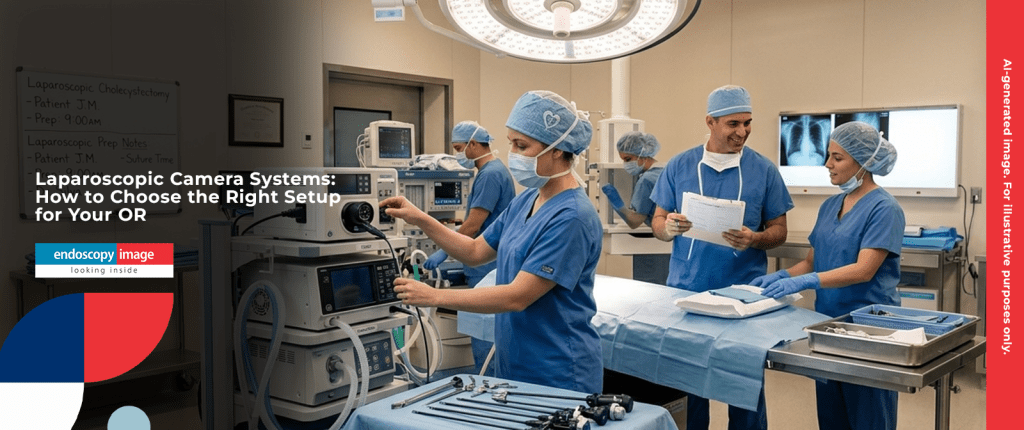
Laparoscopic Camera Systems: How to Choose the Right Setup for Your OR
A laparoscopic camera system is the visual foundation of every minimally invasive surgery. The surgeon’s ability to navigate anatomy, identify structures, and make precise decisions depends directly on the quality and reliability of the imaging system in front of them.
Yet choosing the right system for your operating room is not straightforward. Resolution, light source compatibility, brand ecosystem, procedure types, and budget all play a role — and the differences between available systems are meaningful in practice.
This guide covers the key components of a laparoscopic camera system, the specialties they serve, the leading platforms from Stryker and Karl Storz available through Endoscopy Image, and the practical questions every surgical center administrator should ask before purchasing.
30+Years of experience
2Leading OR brands
7,000+Facilities served worldwide
660+Installations in 2025
Fundamentals
What Is a Laparoscopic Camera System?
A laparoscopic camera system — also referred to as a surgical camera system or endoscopic imaging system for the OR — is the core imaging infrastructure used in minimally invasive procedures. It captures and processes the visual feed from a rigid endoscope or laparoscope and displays it on a surgical monitor for the operating team.
Unlike flexible endoscopy systems used in digestive endoscopy, laparoscopic systems work with rigid instruments inserted through small incisions (trocars). The camera head attaches directly to the eyepiece of the rigid scope and transmits the image to the camera control unit (CCU), which processes and outputs the signal to the monitor.
Core components of a complete laparoscopic imaging setup
1
Camera Control Unit (CCU)
The central processing unit of the imaging system. Receives the raw image signal from the camera head, processes it, and outputs to the monitor. Resolution, color rendering, and image enhancement technologies are determined here.
2
Camera Head
Attaches to the eyepiece of the rigid scope. Contains the image sensor (CCD or CMOS). Must be compatible with the CCU it connects to — camera heads are generally brand- and series-specific.
3
Light Source
Provides illumination through a fiber optic cable connected to the rigid scope. Xenon light sources are the established standard for their color temperature and brightness consistency. LED light sources are increasingly common in newer platforms.
4
Rigid Endoscope / Laparoscope
The optical instrument inserted into the body cavity. Available in different diameters (typically 5mm or 10mm) and viewing angles (0°, 30°, 45°). Must be compatible with the camera head attached to it.
5
Surgical Monitor
Displays the processed image for the surgical team. Medical-grade monitors are designed for the OR environment — appropriate brightness levels, screen coatings, and signal compatibility with the CCU output.
💡 Key principle
A laparoscopic imaging system is only as strong as its weakest component. A 4K camera control unit paired with an outdated light source or a mismatched rigid scope will not deliver its full imaging potential. System components should be selected and verified together.
Applications
Which Surgical Specialties Use Laparoscopic Camera Systems?
Video surgery equipment built around laparoscopic imaging platforms is used across a wide range of minimally invasive surgical specialties. The same camera system can often serve multiple procedure types when paired with the appropriate rigid scope.
🫁
Laparoscopy
Abdominal and pelvic procedures — cholecystectomy, appendectomy, hernia repair, bariatric surgery
🦴
Arthroscopy
Joint visualization and surgery — knee, shoulder, hip, wrist procedures
Endoscopic procedures — ventricular, spinal, and skull base approaches
The breadth of specialties served by modern surgical camera platforms makes them one of the most strategically important equipment investments for any surgical center. A well-chosen system can support multiple OR suites and procedure types across the same facility.
Technical Guide
HD vs. 4K Laparoscopic Imaging: What Actually Changes in the OR
The shift from HD to 4K in surgical camera systems follows the same resolution trajectory as consumer displays — but in the OR, the practical implications go beyond pixel count.
Feature
HD Systems
4K Systems
Resolution
1920 × 1080 pixels
3840 × 2160 pixels (4× HD)
Anatomical detail
Strong for standard laparoscopy
Enhanced fine tissue and vascular detail
Monitor requirement
Compatible with HD surgical monitors
Requires 4K-capable surgical monitor
Typical use case
General MIS, arthroscopy, standard laparoscopy
Complex dissection, high-precision procedures
Secondary market availability
High
Moderate
Example systems
Stryker 1288, 1488, 1588 · Karl Storz IMAGE 1
Stryker 1688 AIM 4K
⚠ Important note
4K resolution requires a compatible 4K surgical monitor to render the full image quality. Installing a 4K camera system on an existing HD monitor will not deliver the resolution benefit. Factor monitor compatibility into your system planning.
Brand Overview
Stryker Surgical Camera Systems
Stryker is one of the most widely adopted brands in minimally invasive surgical imaging globally. Their camera systems are designed for demanding OR environments, with an emphasis on image quality, system integration, and durability across high-volume surgical settings.
All Stryker systems available through Endoscopy Image are pre-owned units sourced through lawful secondary market channels.
System
Resolution
Key Features
Best For
Positioning
Stryker 1688 AIM 4K
4K UHD
Fluorescence Imaging, AutoLight adaptive brightness, full Stryker platform integration
High-complexity laparoscopy, advanced MIS
Advanced
Stryker 1588
Full HD
High color fidelity, precise anatomical detail, robust OR integration
Laparoscopy, arthroscopy, general MIS
High Performance
Stryker 1288
HD
Broad Stryker rigid scope compatibility, multi-specialty versatility, wide installed base
Laparoscopy, arthroscopy, multi-specialty OR
Proven Reference
Stryker 1488
HD
Compact design, reliable imaging, compatible with Stryker rigid endoscopes
The Stryker 1688 AIM 4K includes Fluorescence Imaging capability. This technology, when used with a compatible fluorescent agent and appropriate filter, enables visualization of tissue perfusion and certain anatomical structures that are not readily visible under standard white light. The clinical applicability of fluorescence imaging depends on the procedure type, regulatory clearance in the relevant market, and the specific fluorescent agent used. It is not a standard feature of all laparoscopic procedures.
Brand Overview
Karl Storz Surgical Imaging Systems
Karl Storz has built its reputation on precision optical engineering. The brand is closely associated with high-quality rigid endoscopes and laparoscopes — and their imaging systems are designed to integrate tightly with that optical ecosystem.
KS
Karl Storz IMAGE 1
High-performance endoscopic imaging — multi-specialty OR
Image quality: High-definition imaging with advanced color optimization — precise tissue differentiation for the surgical field
Integration: Designed to work seamlessly with the full Karl Storz rigid endoscope and laparoscope lineup
Specialties supported: Laparoscopy · Urology · Gynecology · Orthopedics · General Surgery
Versatility: Multi-specialty capability makes the IMAGE 1 a strong choice for surgical centers performing procedures across different departments
Karl Storz’s strength lies in the coherence of their system: camera, rigid scope, and light source are engineered to work together. Facilities that invest in Storz rigid scopes benefit most from pairing them with the IMAGE 1 system. Cross-brand mixing (Storz scope with non-Storz camera) may work in some configurations but is not the manufacturer’s recommended setup and can affect image quality.
Comparison
Stryker vs. Karl Storz: A Practical Comparison
Both Stryker and Karl Storz are established leaders in minimally invasive surgical imaging. The right choice for a given facility depends less on brand prestige and more on practical factors: specialty mix, existing equipment, surgeon preference, and procurement budget.
Criterion
Stryker
Karl Storz
Resolution range
HD to 4K UHD (1288 → 1688 AIM)
HD (IMAGE 1)
Fluorescence imaging
Available on 1688 AIM 4K
Not listed in available catalog
Specialty breadth
Laparoscopy, arthroscopy, general MIS
Laparoscopy, urology, gynecology, orthopedics
Rigid scope ecosystem
Broad Stryker scope line
Extensive Storz rigid endoscope catalog
Secondary market availability
Strong
Strong
Best when
Center needs 4K or fluorescence; broad multi-specialty MIS
Center is already in the Storz ecosystem or prioritizes urology/gynecology
💡 Practical note
If your surgical center already has Karl Storz rigid scopes in active use, the IMAGE 1 system is likely the most coherent upgrade path — it is engineered for that ecosystem. If you are building a new OR or expanding into arthroscopy alongside laparoscopy, Stryker’s multi-specialty scope range gives you broader coverage from a single platform.
Decision Guide
Which Laparoscopic Camera System Is Right for Your Surgical Center?
Choose Stryker 1688 AIM 4K
When 4K and fluorescence are clinical priorities
High-complexity laparoscopy, advanced abdominal surgery, centers where enhanced anatomical visualization is part of the surgical protocol. Requires compatible 4K monitor.
Choose Stryker 1588 or 1288
When HD performance and proven versatility are the priority
General laparoscopy, arthroscopy, multi-specialty surgical centers. Strong installed base and secondary market availability. Reliable cost-benefit positioning.
Choose Karl Storz IMAGE 1
When your OR is already in the Storz ecosystem
Facilities with existing Storz rigid scopes in urology, gynecology, or laparoscopy. The IMAGE 1 is optimized for Storz optics and delivers best results in that configuration.
Choose Stryker 1488
When budget efficiency is the primary constraint
Smaller surgical centers, satellite ORs, or facilities adding a second procedure room. Compact, reliable, compatible with the Stryker scope line.
Before You Buy
6 Things to Verify Before Purchasing a Laparoscopic Camera System
Check 01
Confirm camera head and CCU compatibility
Camera heads are typically series-specific. A Stryker 1688 camera head is not interchangeable with a 1288 CCU. Verify the exact pairing before purchasing any component separately.
Check 02
Match the light source to your system
Xenon light sources are the standard for laparoscopy. Confirm the light source type, wattage, and cable compatibility with your rigid scope. A mismatched light source directly affects image brightness and color.
Check 03
Verify monitor compatibility if upgrading to 4K
A 4K CCU requires a 4K surgical monitor to deliver its full resolution. If your OR monitors are HD, upgrading to the Stryker 1688 without also upgrading the monitor will not yield 4K imaging.
Check 04
Check rigid scope compatibility
Rigid scopes (laparoscopes, arthroscopes) from different manufacturers are not always interchangeable at the camera head level. Confirm that your existing or planned rigid scopes are compatible with the camera system you select.
Check 05
Ask about sourcing and equipment history
For any pre-owned system, request documentation of its origin and condition. A reputable supplier will be transparent. If provenance cannot be confirmed, that is a signal to look elsewhere.
Check 06
Plan the full OR tower, not just the camera
A complete laparoscopic OR tower typically includes: CCU + camera head, light source + fiber cable, insufflator (for laparoscopy), monitor(s), and a medical cart. Purchasing components in isolation without planning the full setup creates gaps.
About Us
Endoscopy Image: Pre-Owned Video Surgery Equipment Worldwide
Endoscopy Image, operated by Are Medical LLC and headquartered in Key Biscayne, Miami, Florida, has been supplying hospitals, surgical centers, and clinics with medical equipment for over 30 years. We carry pre-owned video surgery equipment from Stryker and Karl Storz, as well as endoscopy equipment, video gastroscopes, and colonoscopy equipment from Olympus (refurbished), Pentax, and Fujinon.
All equipment we sell consists of pre-owned products acquired through lawful secondary market channels. We operate with full transparency about sourcing and condition — worldwide delivery available.
Looking for a Laparoscopic Camera System for Your OR?
Browse our catalog of pre-owned Stryker and Karl Storz video surgery equipment and contact our team to discuss availability, compatibility, and pricing. Worldwide delivery available.
A complete laparoscopic camera system typically includes a camera control unit (CCU), a camera head, a light source, and a fiber optic cable. For a fully operational OR setup, you also need a compatible rigid endoscope or laparoscope and a surgical monitor. All components must be compatible with each other — camera heads and CCUs are generally brand- and series-specific.
What is the difference between the Stryker 1688 AIM 4K and the Stryker 1288?
The Stryker 1688 AIM 4K is a more recent platform offering 4K UHD resolution and Fluorescence Imaging capability, designed for high-complexity minimally invasive procedures. The Stryker 1288 is an HD camera system with a broad installed base, widely used across laparoscopy, arthroscopy, and general MIS. The 1288 is a proven, cost-effective option; the 1688 is suited for centers where 4K imaging and fluorescence are clinical priorities and a compatible 4K monitor is available.
Can I use a Stryker camera head with a Karl Storz rigid scope?
In some cases, cross-brand adapters exist that allow camera heads from one manufacturer to attach to rigid scopes from another. However, this is not a manufacturer-recommended configuration and may affect image quality, color rendering, or optical performance. For best results, it is generally advisable to keep camera and scope within the same brand ecosystem, or to confirm compatibility specifically with your supplier before purchasing.
Is pre-owned laparoscopic camera equipment a reliable option for surgical centers?
Pre-owned surgical camera systems sourced through verified secondary market channels are used by surgical centers around the world as a cost-effective alternative to new equipment. The key factors are the reliability of the supplier, transparency about the equipment’s origin and condition, and confirmation of functional status prior to purchase. As with any medical device procurement, due diligence on sourcing is essential.
Does Endoscopy Image supply laparoscopic camera systems internationally?
Yes. Endoscopy Image supplies pre-owned video surgery equipment — including Stryker and Karl Storz camera systems — to hospitals, surgical centers, and resellers worldwide. The company is headquartered in Key Biscayne, Miami, Florida. For availability, pricing, and shipping details, contact the team directly at endoscopyimage.com.
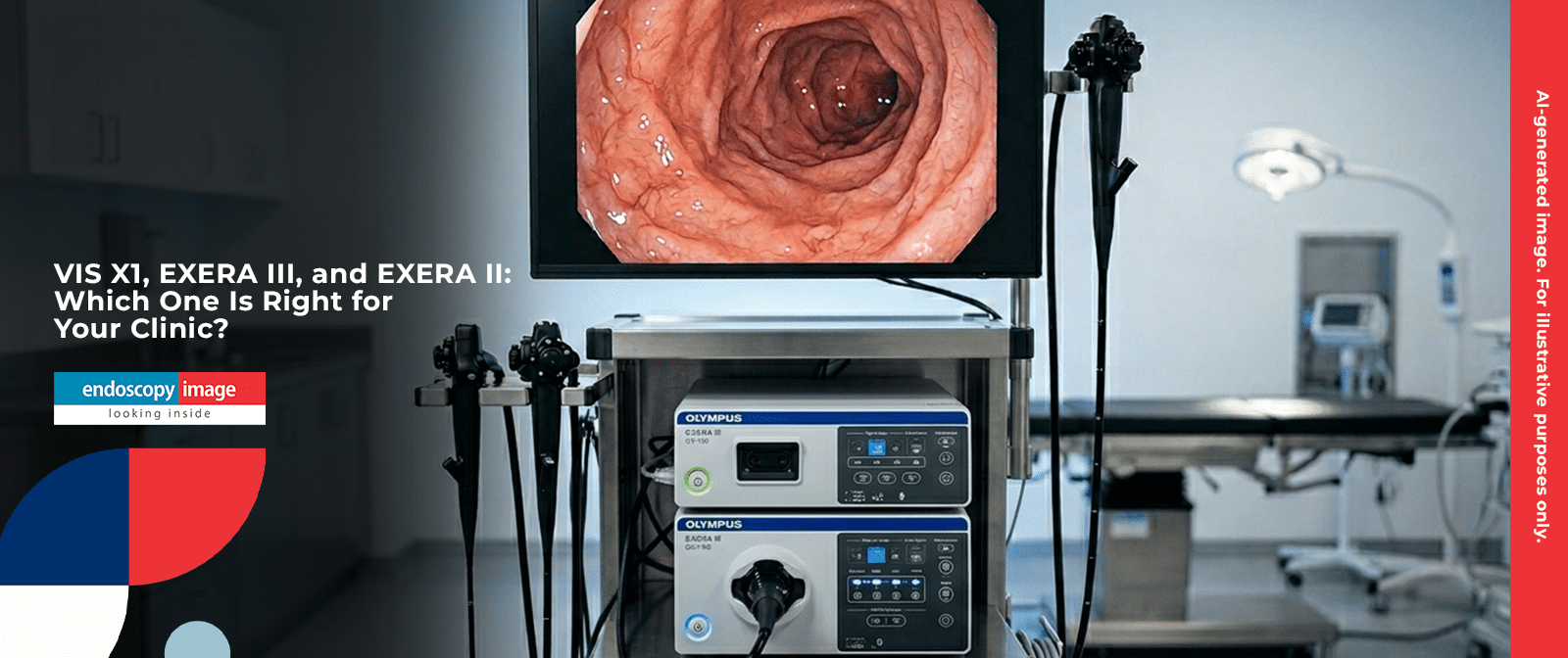
Refurbished Olympus Endoscopy Systems: EVIS X1, EXERA III, and EXERA II — Which One Is Right for Your Clinic?
Olympus is the most widely recognized name in digestive endoscopy worldwide — and for good reason. For decades, the EVIS platform has set the benchmark for image quality, scope ergonomics, and clinical workflow in both upper and lower GI endoscopy.
If you are evaluating a refurbished Olympus endoscopy system for your facility, the most important decision is not just which brand to buy — it is which generation of platform best fits your procedure volume, budget, and clinical requirements.
This guide compares the three Olympus EVIS platforms available through Endoscopy Image: the EVIS X1, the EVIS EXERA III (CV-190), and the EVIS EXERA II (CV-170). All systems are refurbished — pre-owned units sourced through lawful secondary market channels.
30+Years of experience
3Olympus EVIS generations
7,000+Clinics served worldwide
660+Installations in 2025
Brand Overview
Why Olympus Remains the Market Standard in Digestive Endoscopy
In the world of endoscopy equipment, Olympus occupies a unique position. The EVIS platform has gone through continuous evolution since the early days of video endoscopy — and each generation has maintained backward-informed engineering: scopes and processors are designed to work as an integrated system, not as interchangeable components.
This matters in practice. A facility that builds around an Olympus platform benefits from a tightly engineered ecosystem: compatible scopes, consistent image processing, and a large installed base that supports parts availability and technical familiarity.
What makes Olympus platforms consistently reliable
Platform coherence: Processors and scopes are designed together — image processing, illumination, and scope mechanics work as a system
Proven clinical track record: Olympus systems are used in leading hospitals, academic centers, and endoscopy suites globally
NBI (Narrow Band Imaging): Olympus’s proprietary optical contrast technology — available across all three platforms — improves mucosal and vascular visualization
Large installed base: Widespread adoption means strong parts availability and broad technical support in the secondary market
Scope breadth: From standard diagnostic gastroscopes to therapeutic and magnification scopes, each platform supports a full range of procedures
⚠ Important note
All Olympus endoscopy systems sold by Endoscopy Image are refurbished — pre-owned units acquired through lawful secondary market channels. We do not perform in-house repairs or refurbishment. Our role is sourcing, vetting, and connecting buyers with the right systems.
Platform Comparison
The Three Olympus EVIS Platforms: A Side-by-Side Overview
Olympus has developed the EVIS platform through three major generations, each with a different positioning in the market. Here is a direct comparison before going into detail on each.
Latest Generation
EVIS X1
Processor: CV-1500 · Light source: CLV-190
4K UHD resolution
TXI — Texture & Color Enhancement
RDI — Red Dichromatic Imaging
NBI — Narrow Band Imaging
AI-assisted lesion detection
Highest diagnostic capability
Market Reference
EVIS EXERA III
Processor: CV-190 · Light source: CLV-190
Full HD resolution
NBI — Narrow Band Imaging
Compatible with full 190-series scopes
Strong installed base worldwide
Proven in high-volume clinical settings
Best cost-performance balance
Consolidated Value
EVIS EXERA II
Processor: CV-170 · Scopes: GIF-H170, CF-H170
Reliable HD imaging
Proven operational availability
Lower acquisition cost
Suitable for moderate-volume centers
Strong secondary market availability
Straightforward to operate and maintain
Full Feature Comparison Table
Feature
EVIS X1 (CV-1500)
EVIS EXERA III (CV-190)
EVIS EXERA II (CV-170)
Generation
Latest
Current Reference
Consolidated
Resolution
4K UHD
Full HD
HD
NBI
✓ Yes
✓ Yes
✗ No
TXI (Texture & Color Enhancement)
✓ Yes
✗ No
✗ No
RDI (Red Dichromatic Imaging)
✓ Yes
✗ No
✗ No
AI-assisted lesion detection
✓ Yes (supported)
✗ No
✗ No
Compatible gastroscopes
EVIS X1 line
GIF-H190, GIF-HQ190
GIF-H170
Compatible colonoscopes
EVIS X1 line
CF-H190, PCF-H190
CF-H170
Best suited for
Advanced diagnostic & therapeutic endoscopy
High-volume clinical endoscopy
Moderate-volume or budget-sensitive centers
Condition (Endoscopy Image)
Refurbished
Refurbished
Refurbished
Deep Dive
EVIS X1 (CV-1500): The Most Advanced Olympus Platform
The EVIS X1 represents Olympus’s most sophisticated endoscopy platform to date. Built around the CV-1500 video processor, it was designed for high-complexity diagnostic and therapeutic endoscopy environments where image quality and lesion characterization are clinical priorities.
Key technologies
4K UHD resolution: Four times the pixel density of HD, enabling magnification-level detail without a dedicated magnifying endoscope in many cases
TXI — Texture and Color Enhancement Imaging: Enhances surface texture and color rendering of mucosal tissue, improving differentiation of subtle lesions
RDI — Red Dichromatic Imaging: Improves visualization of bleeding sources and hemostasis by enhancing the contrast of red-spectrum tissue
NBI — Narrow Band Imaging: Olympus’s established optical contrast technology for vascular and mucosal pattern analysis
AI support: The EVIS X1 platform includes AI-supported functionalities for endoscopy, as specified by Olympus. Availability and regulatory clearance vary by market and software version.
💡 Who is this for?
The EVIS X1 is a platform suited for high-complexity endoscopy environments where advanced imaging technologies — 4K, TXI, RDI, and NBI — are part of the clinical workflow. As with any medical device, clinical applicability depends on the facility’s protocols, regulatory environment, and procedure types.
Deep Dive
EVIS EXERA III (CV-190): The Market-Reference Platform
If there is one refurbished Olympus endoscopy system that represents the broadest combination of performance, compatibility, and availability in the secondary market, it is the EVIS EXERA III built around the CV-190 processor.
Recognized globally as the standard platform for digestive endoscopy, the CV-190 has been adopted in clinical settings ranging from private outpatient centers to large academic hospitals. Its longevity in the market has created a deep ecosystem of compatible scopes and accessories.
Key features of the EVIS EXERA III
Full HD imaging: Clear, detailed image output for both upper and lower GI endoscopy
NBI — Narrow Band Imaging: Optical contrast technology for vascular and pit pattern analysis — standard on all CV-190 systems
190-series scope compatibility: Pairs with the GIF-H190 and GIF-HQ190 gastroscopes, and the CF-H190 and PCF-H190 colonoscopes
GIF-HQ190 magnification support: When paired with the HQ190 gastroscope, the platform supports optical magnification for detailed early lesion analysis
CLV-190 light source: Designed to work in tandem with the CV-190 for optimized and consistent illumination
✓ Best all-around choice
For most endoscopy centers — from busy outpatient clinics to mid-size hospital units — the EVIS EXERA III (CV-190) delivers the best combination of clinical capability, scope availability, and secondary market value as a refurbished Olympus system.
Deep Dive
EVIS EXERA II (CV-170): Proven Performance at a Lower Investment
The EVIS EXERA II platform — centered on the CV-170 processor — represents an older Olympus generation that remains in active use across a large number of endoscopy facilities worldwide. It is a refurbished endoscopy equipment option that makes strong sense for specific scenarios.
When the EVIS EXERA II is the right call
Clinics establishing a first endoscopy suite with a controlled budget
Centers adding a second procedure room to existing infrastructure
Facilities with lower weekly procedure volume where 4K or advanced optical enhancement is not a clinical priority
Settings where team familiarity with the CV-170 platform already exists
The CV-170 system, combined with the GIF-H170 gastroscope and CF-H170 colonoscope, forms a complete, proven setup for diagnostic upper and lower GI endoscopy. Image quality is reliable HD. The platform is straightforward to operate and has a deep installed base globally.
⚠ One thing to consider
The EVIS EXERA II does not support NBI or advanced optical contrast technologies. For facilities where mucosal pattern analysis is a regular clinical need, the EVIS EXERA III (CV-190) is the more appropriate choice.
Decision Guide
Which Refurbished Olympus System Is Right for Your Clinic?
The right platform depends on four factors: procedure complexity, weekly volume, budget, and whether imaging enhancement technologies (NBI, TXI, AI) are a clinical priority. Here is a practical decision guide.
Choose EVIS X1
For centers where advanced diagnostics are a priority
Busy outpatient clinics, hospital endoscopy units, and multi-room GI suites that need proven HD imaging with NBI and a wide scope ecosystem.
Choose EVIS EXERA II
For budget-sensitive or lower-volume settings
First procedure rooms, satellite clinics, or centers adding capacity without requiring advanced optical imaging enhancement.
Before You Buy
5 Things to Verify When Buying a Refurbished Olympus System
Check 01
Confirm platform generation and processor model
EVIS X1, EXERA III, and EXERA II scopes are NOT cross-compatible. Confirm the exact processor model (CV-1500, CV-190, or CV-170) before selecting scopes.
Check 02
Verify scope condition specifically
For any scope, ask about the condition of the insertion tube, bending section, and working channel — these are the highest-wear components on any endoscope.
Check 03
Confirm the light source is included and compatible
The CLV-190 light source is designed to work with the CV-190 processor. Buying a processor without the matched light source creates immediate compatibility issues.
Check 04
Ask about sourcing and documentation
A reputable supplier will provide documentation of the equipment’s origin. Absence of sourcing transparency is a red flag regardless of how attractive the price appears.
Check 05
Think about the complete system, not just the processor
Processor + matched light source + compatible scopes + monitor = a functional endoscopy setup. Plan the full system before purchasing any individual component.
Check 06
Consider parts and accessory availability
The CV-190 platform has a deeper secondary market than the CV-1500. If long-term parts availability matters, factor this into your platform decision.
About Us
Endoscopy Image: Your Source for Refurbished Olympus Equipment
Endoscopy Image, operated by Are Medical LLC and headquartered in Key Biscayne, Miami, Florida, has been supplying hospitals, clinics, and surgical centers with medical equipment for over 30 years — including 15+ years of operation in the United States.
We carry refurbished Olympus systems across all three EVIS generations, as well as pre-owned video gastroscopes and colonoscopy equipment from the 190-series and 170-series scope lines.
All equipment we sell consists of pre-owned products acquired through lawful secondary market channels. We operate with full transparency about what we source and sell — worldwide delivery available.
Ready to Find the Right Olympus System for Your Facility?
Browse our full catalog of refurbished Olympus endoscopy systems and contact our team to discuss availability, platform compatibility, and pricing. Worldwide delivery available.
What does “refurbished Olympus endoscopy system” mean?
A refurbished Olympus endoscopy system is a pre-owned unit that has been acquired through lawful secondary market channels and made available for resale. At Endoscopy Image, we do not perform in-house repairs or refurbishment. All Olympus equipment we sell consists of pre-owned products sourced through verified secondary market channels — the term “refurbished” reflects their pre-owned status, not a rebuild or repair process.
What is the difference between the Olympus EVIS X1, EXERA III, and EXERA II?
The three platforms represent different generations of the Olympus EVIS endoscopy line. The EVIS X1 (CV-1500) is the most recent generation, featuring 4K UHD resolution, TXI, RDI, NBI, and AI-supported functionalities. The EVIS EXERA III (CV-190) is the widely adopted current-reference platform, offering Full HD imaging with NBI. The EVIS EXERA II (CV-170) is an older generation with reliable HD imaging, suited for centers with moderate volume needs or budget constraints. Scopes are not cross-compatible between platforms.
Are Olympus 190-series scopes compatible with the CV-170 processor?
No. The Olympus 190-series scopes (GIF-H190, GIF-HQ190, CF-H190, PCF-H190) are designed for the EVIS EXERA III platform (CV-190 processor). They are not compatible with the CV-170 processor, which is part of the EVIS EXERA II platform and pairs with the GIF-H170 gastroscope and CF-H170 colonoscope. Always confirm processor-scope compatibility before purchasing any component separately.
Which Olympus system is the best value for a mid-size endoscopy clinic?
For most mid-size clinical endoscopy centers, the EVIS EXERA III (CV-190) represents a strong balance of imaging performance, scope ecosystem breadth, and secondary market availability. It delivers Full HD imaging with NBI and supports a full lineup of 190-series gastroscopes and colonoscopes. The EVIS EXERA II (CV-170) is a viable alternative for centers with lower procedure volume or tighter budgets. The right choice depends on your specific clinical requirements, which we recommend discussing with your procurement team.
Does Endoscopy Image ship Olympus endoscopy equipment internationally?
Yes. Endoscopy Image supplies equipment to hospitals, clinics, surgical centers, and resellers worldwide. The company is headquartered in Key Biscayne, Miami, Florida, and ships to destinations globally. For specific availability, pricing, and shipping information, contact the team directly at endoscopyimage.com.
The three dominant manufacturers in GI endoscopy — Olympus, Pentax Medical, and Fujinon (Fujifilm) — each offer a compelling platform. Each has its flagship imaging technology, its preferred scope ecosystem, and its target facility type. And each has sales teams that will tell you theirs is the best choice.
This article takes a different approach. Rather than reproducing marketing materials, we compare the three platforms on the technical and clinical criteria that actually matter when making a capital equipment decision: imaging modality depth, scope portfolio breadth, light source technology, workflow integration, and the specific facility contexts where each system performs best.
Whether you are equipping a new GI suite, evaluating a system upgrade, or sourcing certified pre-owned endoscopy equipment, this comparison gives you the framework to make an informed decision — independent of vendor positioning.
📌 Scope of This ComparisonThis article focuses on current flagship and mid-tier GI endoscopy platforms from each manufacturer: Olympus EVIS X1 (CV-1500), Pentax EPK-i7010 OPTIVISTA and IMAGINA, and Fujinon ELUXEO VP-7000. Technical information is drawn from manufacturer IFUs, FDA clearance documentation, and official product pages. Where specifications have not been independently verified, this is noted.
The Three Platforms at a Glance
Before going deep into each dimension, here is a top-level overview of the flagship system from each manufacturer currently available in the U.S. and international markets as of 2026.
OLYMPUS
EVIS X1 — CV-1500
Olympus’s most advanced GI platform as of 2026. Flagship of the EVIS line, combining a 5-LED light engine with TXI, NBI, RDI, BAI-MAC, and the new EDOF imaging technology. FDA-cleared in 2023; EZ1500 EDOF scopes cleared May 2025.
PENTAX
EPK-i7010 OPTIVISTA
Pentax’s premium GI processor, featuring i-SCAN digital enhancement and OE (Optical Enhancement) technology — a hybrid digital-optical virtual chromoendoscopy platform. HD+ imaging with freeze-scan technology.
FUJINON
ELUXEO VP-7000 / BL-7000
Fujifilm’s LED-based ELUXEO platform, featuring 4-LED Multi-Light technology with BLI (Blue Light Imaging) and LCI (Linked Color Imaging). The first LED-based endoscopy system commercially introduced in the U.S. (2018), now in its mature generation.
Each manufacturer also offers mid-tier or entry-level systems — Pentax’s IMAGINA, Fujinon’s EPX-4450HD (now a legacy platform), and Olympus’s EXERA III — that remain relevant for facilities with budget constraints or lower procedure volumes. These are addressed in the facility-fit section below.
Imaging Technology: How Each Brand Sees the GI Tract
The most consequential technical difference between the three platforms is their proprietary imaging enhancement technology — the modalities that go beyond standard white light endoscopy. Each brand has developed its own approach to virtual chromoendoscopy, and understanding the differences matters for clinical specialization decisions.
NBI (Narrow Band Imaging): Olympus’s established optical-digital modality, using blue and green wavelengths absorbed by hemoglobin to enhance mucosal and vascular pattern visualization. First introduced globally in 2006; extensively published clinical evidence base.
TXI (Texture and Color Enhancement Imaging): Digital post-processing technology designed to enhance visibility of lesions and polyps by improving color, structure, and brightness of the endoscopic image. Cleared by FDA.
RDI (Red Dichromatic Imaging): Designed to enhance visibility of deep blood vessels and bleeding points — particularly relevant for therapeutic procedures involving GI bleeding.
BAI-MAC (Brightness Adjustment Imaging with Maintenance of Contrast): Corrects brightness in dark areas while maintaining highlight contrast, increasing total distance view.
EDOF (Extended Depth of Field): Olympus’s most recent innovation, using dual prisms to split light into near- and far-focused beams, producing a continuously sharp image across the full depth of field. FDA-cleared May 2025 on EZ1500 series scopes. Available in both GIF-EZ1500 gastroscope and CF-EZ1500DL/I colonoscope.
NBI+TXI combined mode: Launched in Japan November 2025; combines NBI and TXI in a single simultaneous view. International availability pending regulatory approvals.
i-SCAN: Pentax’s digital image enhancement platform, combining three processing algorithms — Surface Enhancement (SE), Contrast Enhancement (CE), and Tone Enhancement (TE). Designed to provide a progressively enhanced view of mucosal texture, surface contour, and vascular structures. Software-based — no modification to the light source required.
OE (Optical Enhancement): An optical-digital hybrid technology available on the EPK-i7010 that filters the light source to provide focused wavelength bands matching hemoglobin absorption characteristics. Combines with i-SCAN to create a three-stage examination workflow: detection, identification, and confirmation.
Twin Mode: Displays HD+ white light and i-SCAN images side-by-side simultaneously, allowing real-time lesion characterization comparison. Designed for teaching environments and complex diagnostic sessions.
IMAGINA i-SCAN: The IMAGINA platform includes a simplified version of i-SCAN with real-time digital enhancement, available in a more affordable system aimed at ASC and outpatient settings.
4-LED Multi-Light Technology: Fujifilm’s foundational innovation for the ELUXEO platform — four independently controlled LEDs (two white, one violet, one green) that enable multiple light combinations beyond white light. First LED-based commercial GI endoscopy system introduced in the U.S. in 2018.
BLI (Blue Light Imaging): Uses short-wavelength blue light to enhance surface structure and vascular patterns, particularly useful for lesion characterization in colonoscopy. Compatible with 700-series and selected 600-series scopes.
LCI (Linked Color Imaging): Enhances mucosal visualization by differentiating the red color spectrum — designed to make the distinction between colorectal lesions and normal mucosa more discernible. Published clinical evidence supports its use in adenoma detection.
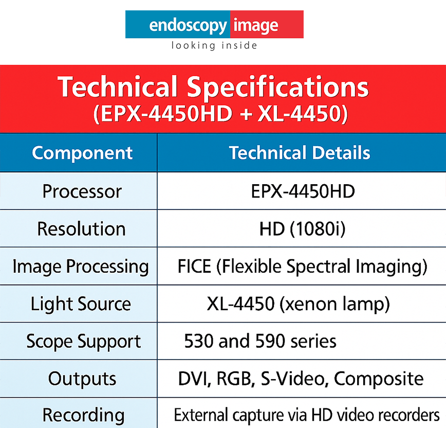
FICE (Flexible Spectral Imaging Color Enhancement): Digital post-processing technology that allows ten configurable settings for virtual chromoendoscopy. Available on both VP-7000 and legacy EPX-4450HD platforms.
BLI-bright mode: A modified BLI mode that increases overall brightness, improving visualization in the proximal colon and other areas with reduced luminal illumination.
Head-to-Head Comparison: Key Technical Parameters
The table below summarizes the most clinically and operationally relevant specifications across the three flagship platforms. Where a feature is platform-specific or generation-dependent, a note is included.
Parameter
Olympus EVIS X1
Pentax EPK-i7010
Fujinon ELUXEO VP-7000
Light Source
5-LED (integrated in CV-1500)
Xenon + OE optical filter
4-LED Multi-Light (BL-7000)
Image Resolution
4K UHD (with compatible monitors)
HD+ (1080p)
Full HD
Proprietary Imaging
TXI, NBI, RDI, BAI-MAC, EDOF
i-SCAN, OE, Twin Mode
BLI, BLI-bright, LCI, FICE
AI Integration
Yes — ENDO-AID CADe (polyp detection)
Not available on EPK-i7010 currently
In development / limited availability
DICOM / EMR Integration
Yes
Yes (endoPRO compatible)
Yes
Touchscreen Interface
Yes (CV-1500 front panel)
Yes (EPK-i7010 and IMAGINA)
Partial / model-dependent
Scope Ergonomics
ErgoGrip — 10% lighter than prev. gen
i10c series — 20–25% lighter scopes
700-series standard ergonomics
Scope Compatibility
EVIS X1 + EXERA III scopes
i10 series; limited backward compat.
700, 600, 500 series (graded compat.)
Entry-level Option
EVIS EXERA III
IMAGINA (ASC-focused)
EPX-4450HD (legacy, still supported)
FDA Clearance (Flagship)
2023 (EVIS X1); EDOF cleared May 2025
FDA-cleared; year varies by model
2017 (VP-7000 / BL-7000)
* Resolution, AI availability, and scope compatibility may vary by configuration, region, and regulatory status. Always confirm with the manufacturer for your specific market. Information sourced from official product pages and FDA documentation as of early 2026.
NBI vs. BLI vs. i-SCAN: Understanding the Imaging Philosophy Differences
The three imaging enhancement platforms reflect fundamentally different technical philosophies — and understanding those differences helps predict which system will integrate better with your clinical workflow.
Olympus NBI: The Most Established Evidence Base
NBI was first introduced commercially in 2006 and has the largest body of peer-reviewed clinical evidence among the three optical enhancement technologies. It modifies the light source output to emit specific blue and green wavelengths that are preferentially absorbed by hemoglobin in mucosal capillaries. The result is enhanced contrast of vascular patterns and mucosal pit structures — particularly useful for Barrett’s esophagus assessment, early gastric cancer detection, and colorectal polyp characterization using validated classification systems (NICE, JNET).
The addition of TXI on the EVIS X1 platform works synergistically with NBI — and the new combined NBI+TXI mode (launched in Japan November 2025, pending international regulatory clearance) is designed to provide mucosal texture and vascular enhancement simultaneously in a single view.
Fujinon BLI and LCI: LED-Native Imaging Enhancement
Fujifilm’s approach with BLI and LCI was designed from the ground up for an LED light source — rather than adapting optical filters to a xenon system. BLI uses short-wavelength blue light to enhance surface and vascular contrast. LCI uses a different LED combination to differentiate the red color spectrum, making subtle differences between inflamed, adenomatous, and normal mucosa more visible to the endoscopist.
A meaningful body of clinical evidence has accumulated around LCI specifically in the context of colorectal adenoma detection rates. Note: while published studies have shown favorable comparisons, this is an active area of research and head-to-head comparisons with NBI are not conclusive as of 2026.
Pentax i-SCAN and OE: Digital-First Flexibility
Pentax’s i-SCAN is entirely software-based — it processes the video signal from the scope without modifying the light source. This means i-SCAN is available across Pentax’s entire compatible scope range, including older i-series instruments, without a hardware upgrade. The three-mode structure (SE, CE, TE) provides progressive examination capability — from surface detail to vascular contrast — within a single imaging pipeline.
The addition of OE (Optical Enhancement) on the EPK-i7010 brings optical filtering into the Pentax workflow for the first time, creating a hybrid digital-optical platform that narrows the gap with Olympus NBI and Fujinon BLI/LCI in terms of light-level enhancement capability. The Twin Mode — simultaneous white light and enhanced imaging on a split screen — is a genuine differentiator for training environments and complex characterization cases.
Light Source Technology: Xenon vs. LED — Why It Matters
The shift from xenon lamp-based to LED-based light sources is one of the most operationally significant transitions in endoscopy over the past decade. Both Olympus and Fujinon have moved their flagship platforms to LED-based light engines. Pentax uses a xenon source on the EPK-i7010 but integrates OE optical filtering; the IMAGINA uses distally-mounted LED lights directly in the scope tip — a different architectural approach.
Factor
LED (Olympus EVIS X1)
LED (Fujinon VP-7000)
Xenon + OE (Pentax EPK-i7010)
Lamp replacement
Not required
Not required
Periodic replacement required
Warmup time
Instant
Instant
Short warmup required
Heat generation
Low
Low
Higher — may affect room temperature
Imaging mode flexibility
Multiple LED combinations enable multi-mode imaging
The IMAGINA system takes a different approach: LEDs are mounted distally — at the tip of the scope itself — rather than in the processor light source. This eliminates the light-guide fiber bundle and, per Pentax’s documentation, provides more uniform illumination at the point of observation. The tradeoff is that these scopes are not backward-compatible with older Pentax processors that rely on light transmission from the proximal end.
Which System Fits Which Facility? A Practical Framework
Beyond specifications, the most useful question is: which system fits the clinical volume, case complexity, and operational constraints of your specific facility? The following framework is based on the publicly documented design intent of each platform, supplemented by the technical parameters above.
Units requiring the most current imaging evidence base
Programs conducting endoscopy research or clinical trials
Facilities where ergonomic fatigue is a concern (ErgoGrip)
PENTAX Best fit for:
Diagnostic and screening-focused GI units
Ambulatory Surgery Centers (ASC) — especially IMAGINA
Training programs that benefit from Twin Mode side-by-side imaging
Facilities seeking lower total cost of ownership
Units replacing older Pentax EPK systems (i-SCAN continuity)
Budget-conscious setups that need genuine HD imaging
FUJINON Best fit for:
Colorectal cancer screening programs (LCI adenoma detection evidence)
Facilities replacing xenon-based Fujinon systems (scope ecosystem continuity)
Mixed-volume hospitals and regional centers
Units that have invested in 600/700-series Fujinon scope inventory
Centers interested in LED-first imaging architecture
Facilities upgrading from EPX-4450HD (direct migration path)
⚠ Important ContextThe “best fit” framework above is based on platform design intent and publicly documented features — not on independent comparative clinical trials between the three systems. No large head-to-head trial comparing patient outcomes across all three platforms under equivalent conditions has been published as of early 2026. Facility decisions should incorporate direct demonstration, IFU review, and service contract evaluation specific to your region.
Scope Ecosystems and Backward Compatibility
A system purchase is not just a processor purchase — it is a commitment to a scope ecosystem. Evaluating backward compatibility and scope portfolio breadth is essential for facilities with existing instrument inventories.
Olympus: The EVIS X1 platform unifies the previously separate EVIS EXERA and EVIS LUCERA scope lines into a single compatible portfolio. This is a significant practical advantage for mixed-inventory units. The newest EZ1500 EDOF scopes require the CV-1500 processor to unlock EDOF functionality.
Pentax: EPK-i7010 is compatible with current i10 series scopes. The IMAGINA uses a distinct i10c scope line with distally-mounted LEDs — these are not backward-compatible with older Pentax processors. Facilities transitioning from EPK-i7000 to EPK-i7010 generally retain scope compatibility.
Fujinon: The VP-7000 offers graded compatibility — 700-series scopes unlock the full BLI/LCI functionality; 600-series scopes offer partial compatibility (BLI/LCI available on selected models); 500-series scopes are compatible for white light and FICE imaging only. The upgrade path is clear and well-documented.
Conclusion: No Universal Winner — But a Right Choice for Your Facility
The comparison between Olympus, Pentax, and Fujinon does not produce a single winner. Each platform is technically capable of supporting high-quality GI endoscopy. What differentiates them are the specific clinical, operational, and economic parameters of your facility.
The Olympus EVIS X1 leads in imaging technology breadth, AI integration, and scope portfolio unification — making it the natural choice for high-complexity, high-volume academic and tertiary programs. The Pentax EPK-i7010 and IMAGINA offer a strong value proposition with genuine imaging enhancement capability and a lower total cost of ownership — particularly suited for ASCs, training environments, and diagnostic-focused units. The Fujinon ELUXEO VP-7000 provides a mature LED-native imaging platform with strong evidence around LCI for colorectal screening, and a clear scope upgrade path for existing Fujinon users.
For facilities evaluating equipment — new or certified pre-owned — across any of these three manufacturers, Endoscopy Image maintains a curated inventory of professionally serviced GI endoscopy systems and can support the technical evaluation process.
Frequently Asked Questions
1. Is Olympus EVIS X1 significantly better than Pentax and Fujinon in image quality?
➡️ This is not a question that can be answered definitively based on available evidence. Each platform offers clinically validated imaging enhancement modes, and image quality in practice depends on the specific scope model, the monitor used, the procedure type, and the endoscopist’s familiarity with the system. EVIS X1 currently has the largest evidence base for its individual technologies (particularly NBI), and its EDOF technology represents a genuinely new optical architecture. However, “best image quality” is not a clinically validated categorical claim for any single platform over the others.
2. Can I use Olympus scopes with a Fujinon or Pentax processor?
➡️ No. Endoscope systems are proprietary — each manufacturer’s scopes use brand-specific connectors, communication protocols, and electrical interfaces. Cross-brand scope-processor combinations are not supported and would violate the IFU of both devices. Facility transitions between brands require a full scope and processor change within the new brand’s ecosystem.
3. What is the cost difference between the three systems?
➡️ None of the three manufacturers publish list prices for their endoscopy systems publicly, and pricing varies significantly by region, configuration, volume discounts, and service contract terms. As a general principle, the Pentax IMAGINA is positioned as the most cost-predictable entry for ASC and outpatient settings. The Olympus EVIS X1 and Fujinon ELUXEO VP-7000 are premium flagship systems with comparable capital investment requirements. Service contract cost and xenon lamp replacement (Pentax EPK-i7010) should be factored into total cost of ownership comparisons.
4. Is it worth buying a refurbished or pre-owned system from any of these three brands?
➡️ Yes — under the right conditions. A certified pre-owned Olympus, Pentax, or Fujinon system from a reputable source, with documented repair history, a passed leak test, and full functional validation, represents a clinically sound investment. The key requirements are: confirmed scope compatibility with the processor, manufacturer or certified repair facility servicing, and verification that the system supports your required imaging modes. Systems from the generation immediately preceding the current flagship — such as Olympus EXERA III, Pentax EPK-i7000, or Fujinon EPX-4450HD — remain fully functional and supported platforms for routine GI procedures.
5. Which system has the best AI integration in 2026?
➡️ As of early 2026, Olympus has the most commercially available AI integration through its ENDO-AID CADe (Computer-Aided Detection) platform, which provides real-time polyp detection assistance during colonoscopy. Fujifilm and Pentax have both indicated AI development roadmaps, but comprehensive commercially available AI-assisted detection systems comparable to ENDO-AID are not yet broadly deployed on the Fujinon or Pentax platforms in the U.S. market as of this writing. This is an actively evolving area.
If you run a gastroenterology clinic or work as a GI specialist, you already know that the terms digestive endoscopy and colonoscopy are not interchangeable — yet confusion between these two procedures costs clinics time, money, and, most importantly, patient outcomes. Knowing exactly when to use each procedure and which equipment best supports your team is the foundation of a high-performing GI practice.
In this guide, we break down the critical differences between upper digestive endoscopy (EGD) and colonoscopy — covering anatomy, clinical indications, procedural workflow, and the specialized equipment each requires. Whether you are evaluating your current endoscopy suite or sourcing new scopes, this article gives you the clinical and operational clarity to make informed decisions.
By the end of this article, you will have a practical framework for matching the right GI procedure to the right patient — and the right equipment to the right procedure.
What Is Digestive Endoscopy? A Clinical Overview
Digestive endoscopy — more precisely, upper gastrointestinal endoscopy or esophagogastroduodenoscopy (EGD) — is a minimally invasive procedure that uses a flexible endoscope to visualize the upper GI tract: the esophagus, stomach, and proximal duodenum. It is one of the most commonly performed procedures in gastroenterology worldwide.
Primary Indications for Upper GI Endoscopy
Persistent heartburn, GERD, or suspected esophageal reflux disease
Dysphagia (difficulty swallowing) or odynophagia
Upper GI bleeding or hematemesis
Suspected peptic ulcer disease or H. pylori infection
Surveillance of Barrett’s esophagus
Evaluation of unexplained weight loss or iron-deficiency anemia
Biopsy of suspicious mucosal lesions in the stomach or duodenum
Equipment Used in Upper Endoscopy
Upper GI endoscopy requires a gastroscope — typically 9–10 mm in diameter — with high-definition imaging capabilities, narrow-band imaging (NBI), and a working channel for biopsy forceps or therapeutic tools. Video processors, light sources, and an insufflation system are also part of a complete endoscopy setup.
Explore our full lineup of endoscopy equipment designed for upper GI procedures.
What Is a Colonoscopy? Scope, Purpose, and Reach
A colonoscopy is a lower GI endoscopic procedure that visualizes the entire large intestine — from the rectum to the cecum — and often the terminal ileum. It is the gold standard for colorectal cancer screening and one of the most impactful preventive procedures in modern medicine.
Primary Indications for Colonoscopy
Colorectal cancer screening (average risk: starting at age 45)
Surveillance after polyp removal or prior colorectal cancer
Lower GI bleeding or hematochezia
Chronic diarrhea or unexplained change in bowel habits
Suspected inflammatory bowel disease (IBD) — Crohn’s or ulcerative colitis
Anemia of unknown origin
Follow-up imaging after abnormal CT colonography
Equipment Used in Colonoscopy
A colonoscope is longer (typically 133–168 cm) and slightly wider than a gastroscope, engineered to navigate the twists of the colon safely. Modern colonoscopes feature variable stiffness, high-definition cameras, and wide-angle optics to maximize adenoma detection rates (ADR). CO₂ insufflation systems, water-jet capabilities, and snares or hemostasis tools round out a colonoscopy suite.
Browse our professional-grade colonoscopy equipment to upgrade your lower GI capabilities.
Digestive Endoscopy vs. Colonoscopy: 6 Key Differences at a Glance
1. Anatomical Reach
Upper endoscopy targets the esophagus, stomach, and duodenum. Colonoscopy targets the rectum, colon, and cecum. Both can reach the terminal ileum when needed for IBD evaluation, though this is more common during colonoscopy.
2. Scope Design and Length
Gastroscopes are shorter (103 cm) and thinner. Colonoscopes are longer (133–168 cm) with variable stiffness sections. Using the wrong scope not only compromises visualization — it can increase procedural risk.
3. Patient Preparation
Upper endoscopy requires only a 6–8 hour fast. Colonoscopy demands a full bowel preparation protocol (low-fiber diet + laxative regimen). Inadequate prep is one of the top reasons for incomplete colonoscopies — making patient education as critical as equipment quality.
4. Sedation and Procedure Duration
EGD typically takes 10–20 minutes with moderate sedation. Colonoscopy averages 20–45 minutes and may require deeper sedation, especially for therapeutic interventions. Anesthesia support and recovery space needs differ accordingly.
5. Therapeutic Capabilities
Both procedures support therapeutic interventions. Upper endoscopy handles band ligation for varices, stent placement, polypectomy, and hemostasis. Colonoscopy enables polyp removal, endoscopic mucosal resection (EMR), tattooing, and stent placement. The therapeutic channel size and accessory compatibility differ between scope types — always verify compatibility before purchasing accessories.
6. Billing and Reimbursement Codes
EGD and colonoscopy carry separate CPT codes with different reimbursement rates. Colonoscopy with polypectomy consistently reimburses at higher rates. Ensuring your documentation and equipment support accurate coding directly impacts your clinic’s financial performance.
When Are Both Procedures Performed Together?
Bidirectional endoscopy — performing both EGD and colonoscopy in the same session — is increasingly common for patients with unexplained anemia, weight loss, or suspected GI bleeding from an unknown source. This approach reduces patient burden (single bowel prep and sedation event) and improves diagnostic yield.
For clinics offering bidirectional endoscopy, having both high-quality gastroscopes and colonoscopes readily available is essential. Endoscopy Image offers both categories of equipment to help you build a complete, efficient GI suite.
Choosing the Right Endoscopy Equipment for Your Practice
Equipment selection is one of the highest-leverage decisions a clinic owner or GI department head can make. The right tools improve adenoma detection rates, reduce procedural complications, and shorten procedure times — all of which affect patient outcomes and practice profitability.
Key Factors to Evaluate
Image quality: HD and 4K video processors dramatically improve mucosal visualization.
Scope compatibility: Ensure scopes are compatible with your existing processors and light sources before purchasing.
Working channel size: Critical for therapeutic procedures — larger channels support bigger accessories.
Reprocessing compatibility: Scopes must be compatible with your reprocessing workflow (HLD or sterilization).
Service and parts availability: Downtime is costly. Choose suppliers with reliable technical support and spare parts.
New vs. refurbished: High-quality refurbished scopes from reputable suppliers offer significant cost savings without compromising performance.
Conclusion: Match the Right Procedure to the Right Patient — and the Right Equipment to Your Practice
Digestive endoscopy and colonoscopy serve distinct clinical purposes, require different equipment, and demand different patient preparation protocols. Mastering the differences between upper GI endoscopy vs. colonoscopy is fundamental for any GI specialist — but equally important is ensuring your practice is equipped with tools that match your procedure volume, patient population, and clinical goals.
At Endoscopy Image, we specialize in helping GI practices, hospitals, and ambulatory surgery centers source the right endoscopy and colonoscopy equipment — from video processors and light sources to scopes and accessories. Whether you are expanding your suite or replacing aging equipment, we have the expertise and inventory to support your clinical mission.
Frequently Asked Questions: Digestive Endoscopy vs. Colonoscopy
1. What is the main difference between digestive endoscopy and colonoscopy?
➡️
The primary difference lies in anatomical reach and clinical purpose. Upper digestive endoscopy (EGD) evaluates the esophagus, stomach, and proximal duodenum, while colonoscopy examines the rectum, colon, and cecum.
Upper endoscopy is commonly indicated for GERD, dysphagia, upper GI bleeding, or suspected peptic ulcer disease. Colonoscopy, on the other hand, is the gold standard for colorectal cancer screening and is frequently used to evaluate lower GI bleeding, chronic diarrhea, or inflammatory bowel disease.
In short, they are complementary procedures targeting different segments of the gastrointestinal tract.
2. How do the equipment requirements differ between upper endoscopy and colonoscopy?
➡️
The procedures require distinct scope designs and supporting systems.
Gastroscopes (used for upper GI endoscopy) are shorter and thinner, typically around 103 cm in length, optimized for upper tract navigation.
Colonoscopes are longer (133–168 cm) and engineered with variable stiffness to safely traverse the colon’s anatomical curves.
In addition, working channel size, accessory compatibility, insufflation systems (air vs. CO₂), and imaging capabilities (HD or 4K processors) must align with the intended procedure. Selecting the wrong equipment can compromise visualization, therapeutic capability, and procedural safety.
3. When should both procedures be performed together?
➡️ Bidirectional endoscopy — performing both EGD and colonoscopy in the same session — is recommended when evaluating:
Unexplained iron-deficiency anemia
Obscure GI bleeding
Significant unintentional weight loss
This approach improves diagnostic yield while reducing patient burden by combining bowel preparation and sedation into a single session. For practices offering this combined strategy, maintaining high-quality equipment for both upper and lower GI procedures is essential for efficiency and clinical accuracy.
Digestive endoscopy has reached a new level of sophistication in recent years. Among the most advanced platforms available today, the Olympus EVIS X1 stands out as a system that goes beyond incremental upgrades, combining advanced imaging, intelligent image processing, and strong clinical focus.
In this article, you will understand what makes the EVIS X1 one of the most advanced endoscopy systems on the market, its key technological differentiators, and in which clinical scenarios it delivers the greatest value.
What Is the Olympus EVIS X1?
The EVIS X1 is Olympus’ next-generation digestive endoscopy platform, designed to significantly improve diagnostic accuracy—especially in the early detection of gastrointestinal lesions.
Unlike previous generations, the EVIS X1 was developed with a strong emphasis on:
Superior image quality
Advanced image-enhancement technologies
Intelligent integration of hardware and software
Direct clinical decision support
Rather than being a simple hardware evolution, the EVIS X1 represents a paradigm shift in endoscopic imaging.
Image Quality: A New Standard in Digestive Endoscopy
One of the most noticeable advances of the EVIS X1 is its significant improvement in image quality. The system delivers sharper images, enhanced contrast, and greater detail of mucosal structures.
In clinical practice, this translates into:
Clearer visualization of vascular patterns
Better definition of lesion margins
Reduced loss of detail in challenging anatomical areas
These improvements directly increase physician confidence during examinations.
Advanced Imaging Technologies That Truly Matter
The EVIS X1 integrates well-established Olympus technologies while introducing new imaging tools designed specifically to support early lesion detection.
🔹 Narrow Band Imaging (NBI)
A widely recognized technology that enhances visualization of superficial vascular patterns and mucosal structures, supporting lesion characterization.
🔹 Texture and Color Enhancement Imaging (TXI)
One of the most important innovations of the EVIS X1. TXI enhances:
Mucosal texture
Color contrast
Subtle differences between healthy and abnormal tissue
In routine clinical use, TXI helps identify abnormalities that may be difficult to detect with standard white-light imaging.
🔹 Enhanced White Light Imaging (WLI)
Even in conventional white-light mode, the EVIS X1 provides noticeable improvements in brightness, contrast, and color fidelity.
Organizations planning long-term investments in endoscopy
While positioned as a premium system, its value lies in diagnostic performance, workflow efficiency, and clinical confidence.
Is the Olympus EVIS X1 Worth the Investment?
From a technical and clinical standpoint, the EVIS X1 is among the most advanced endoscopy systems currently available. However, the decision to invest should consider:
Clinical profile and case mix
Examination volume
Available budget
Medium- and long-term strategic goals
In many cases, investing in the EVIS X1 means raising the diagnostic standard of care, improving clinical outcomes, and strengthening institutional reputation.
Conclusion
The Olympus EVIS X1 is not simply a more modern system—it represents a new generation of digestive endoscopy, focused on precision, intelligent imaging, and real clinical impact.
Endoscopy Image supports physicians, clinics, and hospitals in evaluating whether the EVIS X1 is the right solution for their clinical and operational needs, including new and refurbished options, always with a focus on safety and cost-effectiveness.
FAQ: Olympus EVIS X1 One of the Most Advanced Endoscopy Systems
1.What makes the Olympus EVIS X1 different from previous Olympus endoscopy systems?
➡️ The Olympus EVIS X1 introduces advanced image processing, including Texture and Color Enhancement Imaging (TXI), improved Narrow Band Imaging (NBI), and enhanced white-light imaging. Compared to earlier platforms like the CV-190, it offers superior image clarity, better lesion visualization, and stronger support for early gastrointestinal lesion detection.
2. How does TXI technology improve endoscopy examinations?
➡️ TXI (Texture and Color Enhancement Imaging) enhances subtle differences in mucosal texture and color contrast. This allows physicians to more easily identify early-stage lesions or abnormalities that may be difficult to detect using standard white-light imaging alone, improving diagnostic confidence and accuracy.
3. Is the Olympus EVIS X1 suitable for high-volume clinics and hospitals?
➡️ Yes. The EVIS X1 is designed for high-throughput clinical environments, offering stable performance, compatibility with advanced Olympus endoscopes, and imaging technologies that support consistent diagnostic quality. It is especially suitable for hospitals and clinics focused on screening programs and early cancer detection.
The Pentax EPK-i5000 is a high-definition endoscopy system known for its solid performance and reliable image quality. Designed to serve a wide range of clinical needs, it marked a major step forward in Pentax’s HD imaging technology. In this guide, we’ll explore the key features of the EPK-i5000, its compatible endoscopes, clinical strengths, and when it may be time to upgrade.
What is the EPK-i5000 System?
The EPK-i5000 is a digital video processor introduced by Pentax Medical as part of its i-series HD line. It pairs with a xenon or LED light source and is compatible with 90i and 90K series endoscopes. The system uses CCD sensors and supports digital chromoendoscopy with i-SCAN technology.
Pentax EPK-i5000 Complete Guide: Key Features of the EPK-i5000
High-definition imaging with optimized color reproduction
EG29-i10 / EG29-i10c: HD scopes with advanced maneuverability
EG16-K10: Slim design for pediatric or transnasal procedures
Colonoscopes:
EC34-i10 / EC38-i10: Wide working channels and strong angulation
EC38-K10: Classic scope with stable insertion performance
Technical Specifications (EPX-4450HD + XL-4450)
i-SCAN Technology Explained
Pentax’s i-SCAN digital image enhancement allows clinicians to highlight mucosal surface textures and vascular patterns. It includes three distinct modes: SE (Surface Enhancement), CE (Contrast Enhancement), and TE (Tone Enhancement). This aids in lesion detection and classification, especially during upper GI and colorectal procedures.
When is the EPK-i5000 a Smart Choice?
Ideal for:
Clinics transitioning from standard definition to entry-level HD
Medical centers already using Pentax scopes seeking better image quality
Facilities with trained staff familiar with Pentax ergonomics
Consider upgrading if:
You require full HD (1080p) or 4K video output
You want integrated LED light sources
You plan to incorporate AI-enhanced or digital archiving workflows
Comparison with EPK-i7000 and IMAGINA
Pentax EPK-i5000 Complete Guide: When to Upgrade?
You should consider upgrading from the EPK-i5000 when: – You’re looking for Full HD or integrated LED systems – You need compact solutions for outpatient settings – You’re transitioning to digital imaging and automated report generation
Frequently Asked Questions about the Fujinon EPX-4450HD
1.What endoscopes are compatible with the EPK-i5000 processor?
➡️ The EPK-i5000 supports Pentax’s 90i, 90K, and i10 series endoscopes. This includes both gastroscopes and colonoscopes, such as the EG29-i10, EG16-K10, EC38-i10, and EC38-K10. These scopes offer high-definition imaging and optimized maneuverability for a variety of clinical applications.
2. What are the main imaging advantages of the EPK-i5000 system?
➡️ The system features i-SCAN technology, which enhances visualization with three digital modes: SE (Surface Enhancement), CE (Contrast Enhancement), and TE (Tone Enhancement). These modes help highlight mucosal structures and vascular patterns, improving diagnostic accuracy during GI procedures. routine and advanced procedures.
3. When should a clinic consider upgrading from the EPK-i5000?
➡️ Upgrading is recommended when your facility needs Full HD (1080p) or 4K resolution, integrated LED lighting, or built-in digital archiving. Clinics transitioning into AI-assisted diagnostics or seeking compact, all-in-one solutions may benefit from newer models like the EPK-i7000 or IMAGINA (EPK-i5500c).
Conclusion:
The Pentax EPK-i5000 remains a viable and dependable HD platform for facilities aiming to deliver quality diagnostics without entering the high-cost segment. Its i-SCAN capabilities, robust scope compatibility, and image quality make it a strong choice in 2025. However, for clinics expanding into AI-assisted diagnostics or 4K imaging, newer models may offer a better fit.
Request a Demo or Quote – Fujinon EPX-4450HD
Looking to upgrade or evaluate Pentax systems for your facility? Talk to our specialists and receive a custom quote tailored to your clinical and budgetary needs.
The Fujinon EPX-4450HD is a high-definition endoscopy system from Fujifilm, widely used in diagnostic centers and hospitals. Known for its excellent image quality and advanced features like FICE (Flexible Spectral Imaging Color Enhancement), this system serves as a reliable and cost-effective solution for gastrointestinal procedures. In this guide, we explore its technical capabilities, compatible endoscopes, clinical applications, and when to consider upgrading.
What is the EPX-4450HD System?
The EPX-4450HD is Fujinon’s HD video processor that pairs with the XL-4450 xenon light source. Together, they provide a robust imaging platform for routine and advanced endoscopic diagnostics. The system uses CCD technology and supports both white light and enhanced imaging modes.
Fujinon EPX-4450HD Complete Guide: Key Features of the EPX-4450HD
High-definition output with 1080i resolution
Compatible with Fujinon 590 and 530 series endoscopes
FICE digital chromoendoscopy for enhanced visualization
Adjustable light intensity and automatic brightness control
Video and image recording support via external systems
EG-590WR / EG-590ZW: HD scopes with wide field of view
EG-530WR: Reliable and flexible for general applications
Colonoscopes:
EC-590ZW / EC-590WI: Designed for deeper insertion and therapeutic access
EC-530HL: Slim profile with enhanced maneuverability
Technical Specifications (EPX-4450HD + XL-4450)
FICE Imaging: A Key Differentiator
FICE (Flexible Spectral Imaging Color Enhancement) is Fujinon’s digital image enhancement system that allows clinicians to visualize mucosal patterns and vascular structures with higher contrast and detail. This feature is especially helpful in the early detection of gastrointestinal lesions.
When is the EPX-4450HD a Smart Choice?
Ideal for:
Clinics upgrading from standard definition to HD
Facilities requiring consistent and reliable diagnostic quality
Users familiar with Fujinon’s ergonomics and scope handling
Consider upgrading if:
You need CMOS-based scopes with improved color sensitivity
You require internal image storage or AI-assisted imaging
You’re expanding into advanced therapeutic procedures
Comparison with Newer Fujifilm Systems
Fujinon EPX-4450HD Complete Guide: When to Upgrade?
You should consider upgrading from the EPX-4450HD system when: – You aim to adopt 4K or Full HD systems with LED – You need enhanced image processing tools like BLI or LCI – You’re modernizing infrastructure with integrated storage and AI
Frequently Asked Questions about the Fujinon EPX-4450HD
1.What is the Fujinon EPX-4450HD and what makes it a reliable system in 2025?
➡️ The EPX-4450HD is a high-definition endoscopy video processor developed by Fujifilm. Paired with the XL-4450 xenon light source, it delivers 1080i HD resolution and advanced imaging capabilities like FICE (Flexible Spectral Imaging). In 2025, it remains a cost-effective and dependable platform for routine gastrointestinal diagnostics.
2. What endoscopes are compatible with the EPX-4450HD system?
➡️
This processor supports Fujinon’s 530 and 590 series endoscopes. These include popular models such as:
Gastroscopes: EG-590WR, EG-590ZW, EG-530WR
Colonoscopes: EC-590ZW, EC-590WI, EC-530HL These scopes provide high image clarity, flexibility, and are suitable for both routine and advanced procedures.
3. What are the main imaging advantages of the EPX-4450HD?
➡️ One of its key features is FICE (Flexible Spectral Imaging Color Enhancement), which enhances visualization of mucosal and vascular patterns. This digital chromoendoscopy tool helps in early lesion detection and improves diagnostic accuracy during GI procedures.
Conclusion:
The Fujinon EPX-4450HD remains a trusted HD platform for clinics and hospitals focused on quality diagnostics. Its robust design, FICE imaging, and compatibility with a wide range of scopes make it a strong choice in 2025. For those not yet ready to transition to the VP-7000 series, the EPX-4450HD offers a stable middle ground.
Request a Demo or Quote – Fujinon EPX-4450HD
Need guidance on upgrading or replacing your Fujinon system? Speak to a specialist and receive a custom quote tailored to your needs.