What Makes the Olympus EVIS X1 One of the Most Advanced Endoscopy Systems on the Market
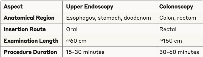
Discover the key differences between endoscopy and colonoscopy procedures. Expert guide for medical..
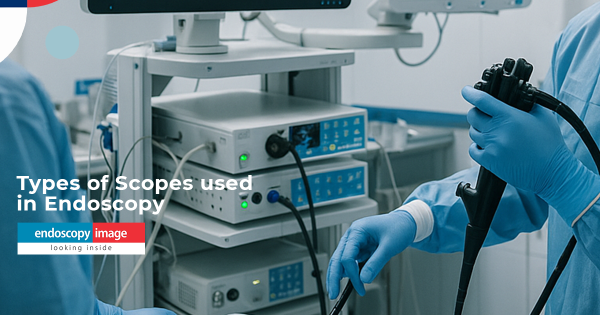
Complete Guide to Next-Generation Digestive Endoscopy Equipment (2026)
Discover the key differences between endoscopy and colonoscopy procedures. Expert guide for medical..
Endoscopy Equipment in Mexico and Latin America: Your Complete 2025 Buying Guide
Discover the key differences between endoscopy and colonoscopy procedures. Expert guide for medical..

Endoscopy and Colonoscopy: The Complete Guide to Understanding Procedure Differences in 2025
Discover the key differences between endoscopy and colonoscopy procedures. Expert guide for medical..